Mitochondria and Aging: The Hidden Engine Behind Energy, Longevity, and Disease
Discover how mitochondrial dysfunction accelerates aging, fatigue, inflammation, and chronic disease. Learn science-backed strategies to boost mitochondrial health, improve energy, and support healthy longevity.
AGING
Dr. T.S. Didwal, M.D.(Internal Medicine)
6/8/202625 min read

How do mitochondria affect aging?
Mitochondria are your cells’ energy factories. As they decline with age, they drive fatigue, muscle loss, brain fog, and inflammation. Key causes include mtDNA mutations, ROS damage, NAD+ loss, and poor mitophagy. You can protect them with Zone 2 exercise, intermittent fasting, polyphenol-rich foods, and targeted supplements like CoQ10 and urolithin A.
Key takeaways :
1. Your mitochondria are your energy and aging clock
These tiny parts of your cells make 90% of your energy. As they wear out, you feel tired, weak, foggy, and age faster. Protecting them is protecting your healthspan.
2. You can’t supplement your way out of a bad lifestyle
Exercise, sleep, and real food do 80% of the work. No pill replaces a 45-minute brisk walk, good sleep, or cutting ultra-processed food.
3. Move your body — it builds new mitochondria
Zone 2 cardio like brisk walking 3–4x per week + 2 days of weights is the most proven way to grow more and better mitochondria. You’ll notice energy in weeks.
4. Give your cells a break with fasting
Going 12–16 hours without food, like 7 pm to 11 am, flips on “cellular cleanup” mode. That clears out damaged mitochondria so healthier ones can take over.
5. Eat for your mitochondria, not against them
Fatty fish, olive oil, berries, leafy greens, nuts, and eggs give mitochondria what they need. Too much sugar, seed oils, and processed snacks damage them.
6. Some supplements help; most are hype
CoQ10 if you’re on statins or 50+, magnesium, and maybe urolithin A have solid data. High-dose vitamin E, resveratrol, and “antioxidant blends” failed human trials and can backfire.
7. Fatigue, brain fog, and slow recovery aren’t “just age”
If you’re constantly tired, weak with exercise, or foggy, your mitochondria may be struggling. Blood tests like GDF15 or lactate can check. Talk to your doctor.
8. It’s never too late — or too early
Mitochondria start declining in your 20s. But studies show 70-year-olds who start Zone 2 + resistance training gain back 20–50% of mitochondrial function in 3 months. Start where you are.
You’re not tired because you’re getting older. You’re tired because your mitochondria are.
Inside every one of your 37 trillion cells sit hundreds to thousands of microscopic engines called mitochondria. Their job: turn food + oxygen into ATP — the energy that powers your brain, heart, and muscles.
But by age 70, you’ve lost roughly 40% of your mitochondrial capacity. MtDNA mutations pile up. The electron transport chain starts “leaking” electrons, flooding cells with damaging ROS. Your body’s cleanup system, mitophagy, slows down. NAD+ — the molecule that keeps mitochondria young — drops by 50% between 20 and 60.
The result? It looks like “normal aging”: fatigue that sleep won’t fix, brain fog, slower recovery, weight gain, weaker grip strength. But it’s not normal. It’s mitochondrial.
And here’s the part medicine rarely tells you: this decline is modifiable.
In human trials, 12 weeks of Zone 2 exercise restored 49% of age-related mitochondrial loss in adults 65+. Fasting for 16 hours triggered mitophagy in muscle within days. Urolithin A, a compound from pomegranates, reactivated mitochondrial recycling in older adults in just 4 months.
This isn’t theory. It’s your biology. And you can change it.
Mitochondria are membrane-bound organelles found in nearly every cell of your body. They evolved from ancient bacteria that were engulfed by early eukaryotic cells more than a billion years ago — which is why they still carry their own unique DNA (mtDNA), entirely separate from the nuclear DNA in your cell's nucleus.
Their primary job is generating adenosine triphosphate (ATP) — the molecule your body uses as currency for virtually every biological process: muscle contraction, nerve signaling, immune responses, DNA repair, hormone synthesis. Without adequate ATP, cellular function degrades rapidly.
But Mitochondria Are Much More Than Energy Factories
Modern biology has revealed a staggering range of mitochondrial functions beyond energy production:
Calcium signaling: Mitochondria regulate intracellular calcium, controlling muscle contraction and neurotransmission
Apoptosis (programmed cell death): They control whether damaged or aged cells undergo orderly self-destruction — a critical anti-cancer mechanism
Reactive oxygen species (ROS) production: As a by-product of energy generation, mitochondria produce free radicals that, in excess, damage DNA and proteins
Heat production (thermogenesis): Brown fat mitochondria generate heat to maintain body temperature
Immune signaling: Mitochondria regulate inflammatory pathways via the NLRP3 inflammasome and other cascades
Stem cell regulation: Mitochondrial metabolism governs whether stem cells remain quiescent or actively divide and repair tissue
The Mitochondria–Aging Connection: What Science Has Discovered
The idea that mitochondria play a central role in aging is not new. The Free Radical Theory of Aging, first proposed by Denham Harman in 1956, suggested that cumulative oxidative damage from mitochondrial ROS drives biological aging. But this was just the beginning.
Decades of research have since revealed a more complex, bidirectional relationship. According to a 2026 review published in Cell Death Discovery by Shan et al., mitochondrial dysfunction and the aging process mutually amplify each other through several overlapping pathways — creating what scientists describe as a "vicious cycle of mitochondrial decline."
"Mitochondrial dysfunction is not merely a consequence of aging — it is now understood as one of its primary drivers, accelerating cellular senescence, tissue degeneration, and systemic inflammation."
— Jia et al., MedComm–Future Medicine, 2025
The Hallmarks of Aging That Involve Mitochondria
The widely cited "Hallmarks of Aging" framework, updated in 2023, identifies twelve core biological processes that drive aging. Mitochondrial dysfunction appears on this list directly — and intersects with at least seven others:
Genomic instability (mtDNA mutations)
Telomere attrition (mitochondria regulate telomerase activity)
Epigenetic alterations (mitochondria supply acetyl-CoA and NAD+ for epigenetic enzymes)
Loss of proteostasis (mitochondrial unfolded protein response)
Deregulated nutrient sensing (AMPK, mTOR, and sirtuins all interface with mitochondria)
Cellular senescence (mitochondrial ROS triggers the senescence-associated secretory phenotype, SASP)
Chronic inflammation ("inflammaging" driven by leaked mtDNA acting as a danger signal)
Key Mechanisms: How Mitochondria Drive Cellular Aging
1. mtDNA Mutations Accumulate Over Time
Unlike nuclear DNA, mitochondrial DNA has limited repair mechanisms and sits very close to the site of ROS production — making it particularly vulnerable to oxidative damage. Over decades, somatic mtDNA mutations accumulate, impairing the efficiency of the electron transport chain (ETC) — the set of protein complexes that actually generate ATP.
Studies in aged humans show that certain tissues (heart, brain, skeletal muscle) accumulate mitochondrial deletions and point mutations at rates that directly correlate with functional decline. By the time we reach our 70s and 80s, a meaningful proportion of mitochondria in these tissues carry mutations that reduce their energy output.
2. The Electron Transport Chain Becomes Leaky
In healthy mitochondria, electrons flow efficiently through the respiratory chain (Complexes I–IV), generating a proton gradient that drives ATP synthesis via Complex V (ATP synthase). In aged or damaged mitochondria, electrons "leak" prematurely to oxygen, forming superoxide radicals — the primary mitochondrial ROS.
This creates a self-perpetuating loop: more ROS → more mtDNA damage → less efficient electron transport → even more ROS.
3. Mitochondrial Membrane Potential Declines
A healthy mitochondrion maintains a strong electrochemical gradient across its inner membrane (around −180 mV). This mitochondrial membrane potential (ΔΨm) is essential for ATP production and for sorting dysfunctional mitochondria for recycling via mitophagy. As we age, ΔΨm declines — a change measurable in blood cells and now being explored as a biomarker of biological age.
4. Mitochondrial Dynamics Are Disrupted
Mitochondria are not static organelles. They constantly undergo fusion (merging together to share contents and buffer damage) and fission (splitting apart to segregate damaged material for disposal). This dynamic balance is regulated by proteins like OPA1, MFN1/2 (fusion) and DRP1, FIS1 (fission).
In aging cells, this balance shifts toward excess fission — producing small, fragmented, dysfunctional mitochondria that are paradoxically less likely to be cleared by mitophagy. This fragmented mitochondrial network is a hallmark of aged tissues.
5. NAD+ Depletion Cripples Mitochondrial Function
NAD+ (nicotinamide adenine dinucleotide) is a critical coenzyme for mitochondrial metabolism and a substrate for sirtuins — the family of proteins that regulate mitochondrial biogenesis, DNA repair, and inflammation. NAD+ levels fall by roughly 50% between age 20 and 60 in human tissues.
Without adequate NAD+, SIRT1 and SIRT3 — key mitochondrial deacetylases — lose activity. PGC-1α, the master regulator of mitochondrial biogenesis (the creation of new mitochondria), is downregulated. The result: fewer new mitochondria are born to replace damaged ones.
Mitophagy: Your Body's Built-In Anti-Aging Recycling System
Mitophagy — the selective autophagy of damaged or dysfunctional mitochondria — is arguably the most powerful natural mechanism your body has to maintain mitochondrial quality. The word comes from the Greek: mito (thread) + phagein (to eat).
Think of mitophagy as a cellular quality control inspector. When a mitochondrion loses membrane potential or becomes damaged beyond repair, the PINK1-PARKIN pathway tags it with ubiquitin chains. Autophagy receptors (NDP52, p62, OPTN) then recruit an autophagosome — a membranous sac — that engulfs the damaged mitochondrion and delivers it to the lysosome for degradation and recycling.
Why Mitophagy Declines With Age
A landmark 2026 review in Cell Death Discovery (Shan et al.) emphasizes that targeting mitochondrial autophagy is one of the most promising anti-aging strategies we currently have. The problem? Mitophagy becomes progressively less efficient with age due to:
Reduced PINK1 expression and PARKIN activity
Declining lysosomal acidity and degradative capacity
Decreased autophagy-initiating ULK1 kinase activity (downstream of mTOR inhibition)
Lower BNIP3L/NIX receptor expression
Impaired mitochondrial fission (which is necessary to "package" mitochondria for mitophagy)
Research Spotlight: Mitophagy and Lifespan
Studies in C. elegans (roundworms), mice, and non-human primates consistently show that enhancing mitophagy extends healthy lifespan. Conversely, animals with impaired PINK1 or PARKIN develop accelerated muscle wasting, neurodegeneration, and metabolic disease — mirroring features of human aging. Human genetic data further supports this link: PINK1 loss-of-function mutations cause early-onset Parkinson's disease, a condition characterized by premature neuronal mitochondrial dysfunction.
How to Stimulate Mitophagy Naturally
Several well-validated interventions are known to upregulate mitophagy:
Caloric restriction or intermittent fasting — reduces mTOR signaling, disinhibiting autophagy
Exercise — particularly endurance and HIIT protocols, which activate AMPK and PGC-1α
Spermidine — a polyamine found in wheat germ and aged cheese, shown to induce autophagy in multiple organisms
Urolithin A — a gut-derived metabolite of pomegranate ellagitannins, shown in human trials to enhance mitophagy in muscle
Rapamycin (mTOR inhibitor) — powerful but prescription-only; used in longevity research protocols
NAD+ precursors (NMN, NR) — restore sirtuin activity that supports mitophagy initiation
Epigenetics, Mitochondria, and the Aging Epigenome
One of the most exciting developments in longevity science is the discovery that mitochondria and the epigenome are in constant communication. The epigenome — the layer of chemical "tags" on your DNA that regulate gene expression — is profoundly sensitive to mitochondrial metabolic output.
A 2025 review in MedComm (An et al.) on epigenetic regulation of aging highlights that mitochondria supply the key metabolites that epigenetic enzymes require:
Acetyl-CoA — substrate for histone acetyltransferases (HATs), which activate gene expression
α-ketoglutarate (α-KG) — required by TET enzymes for DNA demethylation and by Jumonji histone demethylases
NAD+ — substrate for SIRT1, SIRT3, SIRT6 deacetylases, which regulate chromatin compaction
SAM (S-adenosylmethionine) — the universal methyl donor, synthesized via one-carbon metabolism linked to mitochondria
When mitochondria dysfunction, these metabolite pools shift — causing widespread epigenetic dysregulation. The DNA methylation clock (biological age clocks like Horvath's) accelerates. Heterochromatin is lost. Transposons are reactivated. Gene networks governing inflammation and cancer risk are turned on.
"Mitochondria act as metabolic sensors that translate nutritional status and cellular stress into epigenetic programs — effectively deciding how fast or slow an organism ages at the molecular level."
— An et al., MedComm, 2025
Epigenetic Reprogramming: The Frontier of Rejuvenation
Perhaps the most extraordinary new development is partial epigenetic reprogramming — the use of transcription factors (particularly the Yamanaka factors Oct4, Sox2, Klf4) to "reset" the epigenetic age of cells without erasing their identity. Early work in aged mice has shown reversal of visual decline, muscle function, and metabolic disease using this approach. Mitochondrial function recovers in reprogrammed cells — suggesting the epigenetic-mitochondrial axis can be run in reverse. Human clinical trials are expected to begin within 2–4 years.
Signs Your Mitochondria May Be Struggling
Mitochondrial dysfunction rarely announces itself with a single dramatic symptom. Instead, it often manifests as a constellation of vague but persistent complaints — many of which are commonly attributed to "just getting older." Watch for:
Persistent fatigue not explained by poor sleep or anemia — the classic hallmark
Brain fog, difficulty concentrating, or memory lapses
Muscle weakness or rapid fatigue during exercise, especially in muscles with high energy demands
Increased susceptibility to infections (mitochondria regulate immune cell function)
Metabolic slowdown — difficulty losing weight, elevated blood sugar, insulin resistance
Exercise intolerance — heart rate out of proportion to effort, prolonged recovery
Neuropathy or balance issues (neurons are highly mitochondria-dependent)
Elevated blood lactate at rest (a clinical sign of impaired oxidative phosphorylation)
Severe mitochondrial dysfunction can be a feature of primary mitochondrial diseases (rare genetic conditions), chronic illnesses including post-viral syndromes, or medication side effects (notably statins and certain antibiotics). If you experience debilitating fatigue or multi-system symptoms, seek evaluation from a physician — ideally one familiar with metabolic medicine.
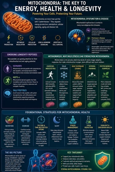
Mitohormesis: Why Mild Stress Can Strengthen Mitochondria
Mitohormesis refers to the phenomenon whereby low levels of cellular stress trigger adaptive responses that ultimately make mitochondria more efficient, resilient, and resistant to aging. Rather than being purely harmful, brief and controlled stressors can stimulate mitochondrial biogenesis, antioxidant defenses, and cellular repair pathways.
Exercise-Induced ROS: Exercise temporarily increases reactive oxygen species (ROS), which act as signaling molecules that activate AMPK, PGC-1α, and endogenous antioxidant systems, ultimately promoting mitochondrial biogenesis and improved metabolic fitness.
Cold Exposure: Short-term cold exposure activates brown adipose tissue, increases mitochondrial density, and stimulates thermogenesis, helping improve metabolic flexibility and energy expenditure.
Heat Exposure (Sauna): Heat stress induces heat-shock proteins (HSPs), enhances cellular repair mechanisms, improves mitochondrial resilience, and may contribute to cardiovascular and longevity benefits observed in regular sauna users.
Hypoxia Training: Intermittent exposure to low-oxygen environments activates hypoxia-inducible factors (HIFs), promoting angiogenesis, mitochondrial adaptation, and improved cellular oxygen utilization.
Phytochemical Hormesis: Plant compounds such as sulforaphane (broccoli), resveratrol (grapes), curcumin (turmeric), and EGCG (green tea) create mild cellular stress that activates protective pathways including Nrf2, sirtuins, and autophagy
.
Mitochondria, Brain Aging, and Neurodegenerative Disease
The brain consumes roughly 20% of the body's energy despite representing only about 2% of body weight, making neurons highly dependent on healthy mitochondria. As mitochondrial function declines with age, the risk of neurodegenerative diseases increases significantly.
Alzheimer's Disease
In Alzheimer's Disease, mitochondrial dysfunction appears years before significant cognitive symptoms develop. Researchers have observed reduced glucose metabolism, impaired ATP production, excessive reactive oxygen species (ROS) generation, and accumulation of damaged mitochondria in affected brain regions. Mitochondrial dysfunction may also promote the formation of amyloid-beta plaques and tau tangles, the pathological hallmarks of Alzheimer's disease. Emerging evidence suggests that improving mitochondrial quality control and mitophagy could help slow disease progression.
Parkinson's Disease
Parkinson's Disease is one of the strongest examples linking mitochondrial dysfunction to neurodegeneration. Mutations in the PINK1 and PARKIN genes—both critical regulators of mitophagy—cause familial forms of Parkinson's disease. Dysfunctional mitochondria accumulate within dopamine-producing neurons, leading to oxidative stress, impaired energy production, and ultimately neuronal death. These findings have made mitochondrial-targeted therapies a major focus of Parkinson's research.
Amyotrophic Lateral Sclerosis (ALS)
In Amyotrophic Lateral Sclerosis, damaged mitochondria are frequently observed within motor neurons and skeletal muscle. Mitochondrial abnormalities contribute to impaired energy metabolism, excessive oxidative stress, calcium dysregulation, and neuroinflammation. These changes may accelerate the progressive loss of motor neurons that characterizes ALS. Although no mitochondrial therapy has yet proven curative, several experimental treatments aim to improve mitochondrial function and reduce oxidative damage.
Key Takeaway
A growing body of evidence suggests that mitochondrial dysfunction is not merely a consequence of neurodegeneration—it may be an early driver of neuronal aging and disease progression. Maintaining mitochondrial health through regular exercise, metabolic health optimization, adequate sleep, and strategies that support mitophagy may therefore represent an important approach to preserving long-term brain function and cognitive resilience.
Diet Strategies to Support Mitochondrial Health and Slow Aging
The single most powerful dietary lever for mitochondrial health is managing energy intake and meal timing. Here's what the evidence supports:
Intermittent Fasting and Time-Restricted Eating
Periods of fasting activate AMPK (the cellular energy sensor), suppress mTOR (the growth-promoting kinase that inhibits autophagy), and upregulate PGC-1α — all of which promote mitophagy and mitochondrial biogenesis. A 2023 meta-analysis found that time-restricted eating (eating within an 8–10 hour window) significantly improved metabolic markers associated with mitochondrial efficiency, including fasting insulin and inflammatory markers.
Practical approach: A 16:8 fasting protocol (16 hours fasting, 8-hour eating window) is achievable for most people and has the broadest evidence base. Start with 12:12 and extend gradually.
The Mitochondria-Supportive Plate
Prioritize these nutrient categories:
Polyphenols (berries, dark chocolate, green tea, olive oil) — activate sirtuins and Nrf2 pathways that upregulate antioxidant defenses and mitophagy
Omega-3 fatty acids (fatty fish, walnuts, flaxseed) — incorporated into mitochondrial inner membranes, improving fluidity and reducing ROS
Magnesium-rich foods (spinach, almonds, avocado) — essential cofactor for over 300 mitochondrial enzymes including ATP synthase
B-vitamins (especially B1, B2, B3, B5) — directly required as coenzymes in the Krebs cycle and electron transport chain
CoQ10-rich foods (organ meats, sardines, beef) — CoQ10 is the electron shuttle between Complexes I/II and III
Cruciferous vegetables (broccoli, Brussels sprouts) — sulforaphane activates Nrf2, boosting mitochondrial antioxidant enzymes
Pomegranate — ellagitannins converted by gut bacteria to urolithin A, a potent mitophagy inducer
Foods to Minimize
Ultra-processed foods (drive neuroinflammation and gut dysbiosis that impairs mitochondrial signaling)
Excessive refined sugars (cause glycation of mitochondrial proteins)
Industrial seed oils high in linoleic acid (may impair mitochondrial membrane fluidity when consumed in excess)
Chronic heavy alcohol consumption (directly toxic to mitochondrial membranes and ETC complexes)
Sample Day: Mitochondria-Supportive Eating Pattern
Eating window: 11 AM – 7 PM (16:8 fast)
11 AM – Breakfast: 2 eggs + smoked salmon + avocado + spinach + green tea
2 PM – Lunch: Grilled sardines or mackerel, large mixed salad with extra virgin olive oil, walnuts, blueberries
5 PM – Snack: Dark chocolate (85%+), handful of almonds, small portion of pomegranate seeds
7 PM – Dinner: Pasture-raised chicken or beef liver, roasted broccoli + Brussels sprouts, sweet potato
Exercise: The Most Powerful Mitochondrial Biogenesis Signal Known
If there is one intervention with the strongest, most consistent evidence for improving mitochondrial health across the lifespan, it is physical exercise. And the mechanism is well understood: exercise creates a temporary energy deficit that activates AMPK, which in turn triggers PGC-1α — the master transcriptional coactivator of mitochondrial biogenesis.
When PGC-1α is activated, cells begin building new mitochondria, increasing their number, size, and efficiency. Regular exercise also enhances mitophagy, improving the turnover of damaged mitochondria. According to a 2024 review in Aging and Disease (Phua et al.), exercise remains the most accessible and effective "mitochondrial rejuvenation tool" currently available.
Optimal Exercise Protocol for Mitochondrial Health
Here is the exercise breakdown rewritten into a highly skimmable, structured points format:
1. Endurance Training (Zone 2)
Exercise Type: Moderate-intensity aerobic movement (e.g., cycling, brisk walking, jogging).
Frequency: 3–4 times per week.
Duration: 45–60 minutes per session.
Primary Mechanism: Drives maximal PGC-1α activation (the master regulator of mitochondrial biogenesis), induces mitophagy (cellular cleanup of damaged mitochondria), and optimizes fat oxidation.
Evidence Level: Very Strong (backed by multiple Randomized Controlled Trials).
2. High-Intensity Interval Training (HIIT)
Exercise Type: Short, intense bursts of effort (e.g., 4 intervals of 4 minutes at approximately 90% maximum heart rate).
Frequency: 2 times per week.
Duration: 20–30 minutes per session.
Primary Mechanism: Activates the AMPK pathway, suppresses mTOR expression, and rapidly increases total mitochondrial density.
Evidence Level: Strong (backed by Randomized Controlled Trials).
3. Resistance Training
Exercise Type: Compound lifts, bodyweight exercises, or weight machines.
Frequency: 2–3 times per week.
Duration: 30–45 minutes per session.
Primary Mechanism: Stimulates muscular mitochondrial biogenesis, optimizes the balance between IGF-1 and mTORC1 pathways, and actively prevents sarcopenia (age-related muscle loss).
Evidence Level: Strong.
4. Yoga / Tai Chi
Exercise Type: Low-intensity mindful movement paired with targeted stress reduction.
Frequency: 2–3 times per week.
Duration: 30–60 minutes per session.
Primary Mechanism: Reduces chronic cortisol levels (elevated stress hormones are known to actively impair mitophagy) and restores parasympathetic nervous system balance.
Evidence Level: Moderate.
Zone 2 training deserves special attention. Research by exercise physiologist Iñigo San Millán has popularized Zone 2 as the "metabolic sweet spot" for mitochondrial health — the intensity at which you can maintain a conversation, but would struggle to sing. At this intensity, Type I slow-twitch muscle fibers are maximally recruited, forcing mitochondria to oxidize fat for fuel and generating the most potent PGC-1α signaling.
A Practical Weekly Schedule
Monday: 45 min Zone 2 (cycling or brisk walking)
Tuesday: 30–40 min resistance training (compound movements)
Wednesday: Active recovery (yoga, walking, stretching)
Thursday: HIIT — 4×4 min at high intensity, 3 min recovery between sets
Friday: 30 min resistance training
Saturday: 60 min Zone 2 (longer aerobic session)
Sunday: Rest or gentle movement
Supplements and Emerging Therapies for Mitochondrial Ageing
The supplement landscape for mitochondrial health is crowded with hype. Here's a grounded look at what the evidence actually supports — from strong to speculative.
Tier 1: Strong Evidence
CoQ10 / Ubiquinol: Coenzyme Q10 is the mobile electron carrier in the ETC, essential for Complex III activity and serving as a membrane antioxidant. Production declines with age and is further suppressed by statins. Supplementation (100–300 mg/day) consistently reduces oxidative stress markers and may improve energy in statin users and those over 50. Ubiquinol (the reduced form) has superior absorption.
NMN / NR (NAD+ Precursors): Both nicotinamide mononucleotide (NMN) and nicotinamide riboside (NR) raise tissue NAD+ levels in humans, with NMN showing particularly robust data in a 2022 RCT in healthy older adults. Raising NAD+ restores sirtuin activity, supports PARP-dependent DNA repair, and enhances PGC-1α — all of which benefit mitochondrial quality.
Magnesium: Deficiency is epidemic (up to 50% of Western adults), and magnesium is a required cofactor for mitochondrial ATP synthesis, Complex I/II activity, and membrane potential maintenance. Magnesium glycinate or malate are the best-absorbed forms.
Tier 2: Promising Evidence
Urolithin A: A 2022 human trial published in Nature Aging showed oral urolithin A (500–1000 mg/day) significantly enhanced mitophagy markers and mitochondrial gene expression in skeletal muscle of older adults — the first human trial to demonstrate direct mitophagy induction by a supplement. Follow-up trials are ongoing.
Alpha-lipoic acid (ALA): A mitochondria-derived antioxidant and cofactor for Krebs cycle enzymes (pyruvate dehydrogenase, α-KG dehydrogenase). R-ALA is the biologically active form. Evidence supports its role in reducing mitochondrial oxidative stress, particularly in the context of diabetic neuropathy.
Spermidine: This naturally occurring polyamine (found in wheat germ, mushrooms, soybeans, aged cheese) is one of the most studied autophagy inducers. Epidemiological data and animal trials show associations between higher spermidine intake and reduced cardiovascular mortality and improved cognitive aging. A 2021 randomized trial showed cognitive improvements in older adults with subjective memory complaints.
PQQ (Pyrroloquinoline quinone): Acts as a mitochondrial cofactor and has shown ability to stimulate mitochondrial biogenesis (PGC-1α activation) in preliminary human studies at doses of 20 mg/day. Evidence is still limited but early results are encouraging.
Tier 3: Emerging / Experimental
Rapamycin: The mTOR inhibitor with perhaps the most dramatic lifespan-extending effects in mouse models. Some longevity physicians are prescribing low-dose, intermittent rapamycin off-label based on compelling animal data. Human RCT data remain limited. Risks include immunosuppression and metabolic effects. Not recommended without physician supervision.
SS-31 (Elamipretide): A mitochondria-targeted peptide that specifically concentrates in the inner mitochondrial membrane and stabilizes cardiolipin — a lipid essential for ETC Complex I/III/IV supercomplexes. Phase 2 trials show promise in heart failure and frailty. Not yet commercially available.
Mitochondria-targeted antioxidants (MitoQ, SkQ1): These molecules link antioxidants (ubiquinone or plastoquinone) to a lipophilic cation that concentrates 500-fold inside mitochondria. Promising in animal models; limited but early positive human data
Many supplements interact with medications and underlying health conditions. NMN and NR may affect insulin sensitivity; CoQ10 can interact with anticoagulants; alpha-lipoic acid affects blood glucose. Always discuss supplementation with your healthcare provider, especially if you are managing a chronic condition.
Mitochondrial-Derived Peptides: An Emerging Frontier in Longevity Research
Beyond producing energy, mitochondria also generate small signaling molecules known as mitochondrial-derived peptides (MDPs). These peptides act as cellular messengers that help regulate metabolism, stress resistance, inflammation, and healthy aging. Three of the most studied MDPs are Humanin, MOTS-c, and SHLPs.
Humanin
Humanin was the first mitochondrial-derived peptide discovered. It appears to protect cells against oxidative stress, apoptosis (programmed cell death), and age-related cellular damage. Laboratory studies suggest Humanin may protect neurons from amyloid-beta toxicity, making it a potential target in Alzheimer's disease research. Humanin levels tend to decline with age, and lower concentrations have been associated with metabolic dysfunction and increased cardiovascular risk.
MOTS-c
MOTS-c (Mitochondrial Open Reading Frame of the 12S rRNA-c) is often described as an "exercise-mimetic" peptide because it influences many of the same metabolic pathways activated by physical activity. MOTS-c enhances insulin sensitivity, promotes glucose utilization, activates AMPK, and improves metabolic flexibility. Animal studies have shown improvements in physical performance and metabolic health, leading researchers to investigate its potential role in obesity, type 2 diabetes, and healthy aging.
SHLP Peptids
Small Humanin-Like Peptides (SHLP1–6) are a family of mitochondrial-derived peptides structurally related to Humanin. Early research suggests they may regulate mitochondrial metabolism, reduce oxidative stress, improve insulin signaling, and support cellular survival. Some SHLPs appear to have anti-inflammatory and cytoprotective effects, although human data remain limited.
Why They Matter for Aging
Mitochondrial-derived peptides represent a new layer of communication between mitochondria and the rest of the cell. Rather than merely serving as power generators, mitochondria appear to function as endocrine-like signaling organelles that influence whole-body aging. Although Humanin, MOTS-c, and SHLPs have shown promising effects in animal and laboratory studies, large human clinical trials are still needed before these peptides can be considered established anti-aging therapies.
Bottom line: Humanin, MOTS-c, and SHLP peptides are among the most exciting emerging areas in longevity science, offering a potential link between mitochondrial health, metabolic resilience, and healthy aging. However, their clinical applications remain experimental at present.
Evidence Summary: Key Studies at a Glance
Mitophagy Targeting (Shan et al., 2026; Cell Death Discovery)
Demonstrated that impaired mitophagy is a major contributor to aging.
Enhancing mitochondrial autophagy extended lifespan and improved healthspan in experimental models.
Population: Animal and human review data.
Evidence Quality: High.
Mitochondrial Therapies (Jia et al., 2025; MedComm – Future Medicine)
Identified multiple therapeutic targets for age-related mitochondrial dysfunction.
Highlighted precision medicine approaches aimed at restoring mitochondrial health.
Population: Comprehensive scientific review.
Evidence Quality: High.
Exercise as a Rejuvenation Tool (Phua et al., 2024; Aging and Disease)
Exercise increased mitochondrial biogenesis and enhanced mitophagy.
Associated with improvements in metabolic health and reductions in biological aging markers.
Population: Review of human randomized controlled trials.
Evidence Quality: High.
Epigenetic Reprogramming (An et al., 2025; MedComm)
Showed that age-related epigenetic alterations linked to mitochondrial dysfunction may be reversible.
Partial cellular reprogramming demonstrated rejuvenation effects in experimental models.
Population: Animal and laboratory studies.
Evidence Quality: Moderate-High.
Urolithin A Supplementation (Liu et al., 2022; Nature Aging)
Increased expression of mitophagy-related genes.
Improved markers of mitochondrial function in skeletal muscle of older adults.
Population: Randomized controlled trial (n = 66, age 65+).
Evidence Quality: Moderate-High.
NMN Supplementation (Igarashi et al., 2022; NPJ Aging)
Increased blood NAD+ levels by approximately 38%.
Improved gait speed, handgrip strength, and sleep quality compared with placebo.
Population: Randomized controlled trial (n = 42, age 65+).
Evidence Quality: Moderate.
Spermidine Supplementation (Wirth et al., 2021; Cortex)
Improved memory performance after 3 months of supplementation.
Benefits observed in older adults with subjective cognitive decline.
Population: Randomized controlled trial (n = 85, age 60–80).
Evidence Quality: Moderate.
HIIT vs. Moderate Exercise (Robinson et al., 2017; Cell Metabolism)
High-intensity interval training (HIIT) reversed age-related declines in mitochondrial capacity more effectively than resistance training.
Older adults experienced a 49% improvement in mitochondrial function compared with 19% from resistance training alone.
Population: Randomized controlled trial (n = 72, age 18–80).
Evidence Quality: High.
Strength of Evidence for Mitochondrial Longevity Interventions
Exercise
Mechanistic evidence: Very strong
Human biomarker evidence: Very strong
Lifespan evidence: Indirect but strong
Key takeaway: Exercise remains the most proven intervention for improving mitochondrial function, stimulating mitophagy, and promoting healthy aging.
Intermittent Fasting
Mechanistic evidence: Strong
Human biomarker evidence: Moderate
Lifespan evidence: Limited in humans
Key takeaway: Fasting activates autophagy and metabolic repair pathways, but direct human lifespan data are still lacking.
Urolithin A
Mechanistic evidence: Strong
Human biomarker evidence: Moderate
Lifespan evidence: None currently in humans
Key takeaway: One of the most promising mitophagy-enhancing compounds, with encouraging clinical data for mitochondrial health.
NMN (Nicotinamide Mononucleotide)
Mechanistic evidence: Strong
Human biomarker evidence: Moderate
Lifespan evidence: None currently in humans
Key takeaway: NMN reliably raises NAD+ levels and may improve aspects of physical function, but lifespan benefits remain unproven.
Bottom Line
Strongest overall evidence: Exercise
Most promising lifestyle strategy: Intermittent fasting
Most promising mitophagy-targeting supplement: Urolithin A
Most promising NAD+-boosting intervention: NMN
Current reality: Lifestyle interventions have far stronger evidence than any supplement currently available.Overall Conclusion
Across the current evidence base, exercise, mitophagy enhancement, NAD+ restoration, and metabolic interventions consistently emerge as the most promising strategies for maintaining mitochondrial function and promoting healthy aging. While supplements such as urolithin A, NMN, and spermidine show encouraging results, lifestyle interventions remain the most strongly supported and clinically actionable approaches.
Common Myths and Mistakes About Mitochondrial Health
Myth 1: "Taking antioxidants will fix mitochondrial damage"
The relationship between antioxidants and mitochondria is more nuanced than supplement marketing suggests. Mitochondrial ROS at low levels serve important signaling functions — triggering adaptive stress responses (hormesis) that actually strengthen mitochondria. Excessive antioxidant supplementation (particularly isolated vitamin C or E in high doses) can blunt these beneficial signals. Whole-food polyphenols, which activate the body's own antioxidant systems (Nrf2, glutathione), are preferable to mega-dose antioxidant pills.
Myth 2: "Only old people need to think about this"
Mitochondrial DNA mutations begin accumulating in your 20s. Exercise capacity (VO₂max) — a proxy for mitochondrial fitness — peaks in your late 20s to early 30s and declines by roughly 1% per year without intervention. The protective habits built in your 30s and 40s compound dramatically; waiting until symptoms appear is a costly delay.
Myth 3: "More exercise is always better for mitochondria"
Overtraining without adequate recovery increases chronic oxidative stress and impairs mitophagy. The beneficial effects of exercise on mitochondria are achieved through the recovery phase, not the workout itself. Sleep, nutrition, and scheduled rest are as important as the exercise stimulus.
Myth 4: "If you have mitochondrial problems, supplements will save you"
No supplement can compensate for a sedentary lifestyle, poor sleep, chronic stress, or a diet high in ultra-processed foods. Supplements are adjuncts to lifestyle — not replacements. The hierarchy is clear: sleep → movement → nutrition → stress management → targeted supplementation.
Myth 5: "Mitochondrial supplements are all the same"
Quality, form, and dosing vary enormously. NMN versus NR have different pharmacokinetics; ubiquinol versus CoQ10 differs in absorption; R-ALA versus S-ALA has opposing stereochemistry effects. Research the specific form used in the studies, not just the generic supplement name.
Frequently Asked Questions
What is the connection between mitochondria and aging?
Mitochondria are responsible for producing most of the energy (ATP) that powers cellular function. As we age, mitochondria accumulate DNA damage, generate more oxidative stress, and become less efficient at producing energy. This decline contributes to many hallmarks of aging, including inflammation, cellular senescence, metabolic dysfunction, and reduced tissue repair capacity. As a result, mitochondrial dysfunction is increasingly viewed as both a driver and a consequence of biological aging.
Can you reverse mitochondrial aging?
Completely reversing mitochondrial aging is not currently possible, but research suggests that aspects of mitochondrial function can be improved. Regular exercise, healthy nutrition, adequate sleep, caloric moderation, and emerging interventions such as NAD+ precursors and mitophagy-enhancing compounds may help restore mitochondrial efficiency, stimulate mitochondrial biogenesis, and improve cellular resilience.
What is mitophagy and why is it important for longevity?
Mitophagy is the selective removal and recycling of damaged or dysfunctional mitochondria. It acts as a cellular quality-control system that prevents the accumulation of defective mitochondria that can generate excessive oxidative stress and inflammation. Efficient mitophagy helps maintain a healthy mitochondrial population and is strongly associated with healthy aging and increased lifespan in multiple experimental models.
Which exercises are best for mitochondrial health?
Both aerobic and resistance exercise support mitochondrial health. Zone 2 endurance training is particularly effective at stimulating mitochondrial biogenesis and improving fat oxidation, while high-intensity interval training (HIIT) enhances mitochondrial capacity and metabolic flexibility. Resistance training also improves mitochondrial function while preserving muscle mass, making a combination of all three approaches ideal.
What are the best supplements for mitochondrial health?
The strongest evidence currently supports CoQ10, magnesium, and NAD+ precursors such as NMN and nicotinamide riboside (NR). Emerging compounds including urolithin A, spermidine, alpha-lipoic acid, and PQQ show promise for enhancing mitophagy and mitochondrial function. However, supplements should complement—not replace—exercise, sleep, and healthy nutrition.
How does diet affect mitochondrial function?
Diet directly influences mitochondrial metabolism, oxidative stress, and cellular signaling pathways. Nutrient-dense foods rich in polyphenols, omega-3 fatty acids, vitamins, and minerals support mitochondrial efficiency, while excessive consumption of ultra-processed foods, refined sugars, and chronic overnutrition can impair mitochondrial function and accelerate aging-related damage.
Does intermittent fasting improve mitochondrial health?
Yes. Intermittent fasting and time-restricted eating activate energy-sensing pathways such as AMPK while reducing mTOR activity. These changes promote autophagy, mitophagy, mitochondrial biogenesis, and metabolic flexibility. Research suggests fasting may improve several biomarkers associated with mitochondrial function and healthy aging.
What is the role of NAD+ in mitochondrial aging?
NAD+ is a critical coenzyme involved in cellular energy production and mitochondrial metabolism. It also activates sirtuins, proteins that regulate mitochondrial biogenesis, DNA repair, and stress resistance. Because NAD+ levels decline with age, mitochondrial efficiency often decreases. Maintaining healthy NAD+ levels may help support mitochondrial function and cellular health during aging.
Can poor sleep damage mitochondria?
Yes. Chronic sleep deprivation increases oxidative stress, disrupts mitochondrial energy production, impairs cellular repair mechanisms, and promotes inflammation. Over time, inadequate sleep can contribute to mitochondrial dysfunction, metabolic disturbances, cognitive decline, and accelerated biological aging. Consistent, high-quality sleep remains one of the most important pillars of mitochondrial health.
Are mitochondrial diseases the same as age-related mitochondrial dysfunction?
No. Mitochondrial diseases are usually rare genetic disorders caused by inherited mutations affecting mitochondrial function. Age-related mitochondrial dysfunction, by contrast, develops gradually over time due to accumulated damage, oxidative stress, declining mitophagy, and metabolic changes. While both involve impaired mitochondrial function, their causes, severity, and clinical presentation differ substantially.
The Takeaway: Your Mitochondria Are Not Your Fate
The science is unambiguous: mitochondria and aging are deeply and causally intertwined. As mitochondrial quality declines — through ROS-driven mtDNA mutations, failing mitophagy, NAD+ depletion, and disrupted dynamics — the biological hallmarks of aging accelerate across virtually every organ system.
But here is what's remarkable: this process is not simply inevitable. It is, to a significant and measurable degree, modifiable. The interventions with the strongest evidence — regular aerobic and resistance exercise, time-restricted eating, whole-food nutrition rich in polyphenols and omega-3s, quality sleep — directly target mitochondrial biology. They are free, accessible, and their benefits compound over decades.
Beyond lifestyle, emerging research into mitophagy enhancement (urolithin A), NAD+ restoration (NMN/NR), epigenetic reprogramming, and mitochondria-targeted peptides represents an extraordinary frontier. The next decade of longevity science will very likely be defined by our ability to intervene at the mitochondrial level with precision.
Your Action Plan — Start This Week
Begin Zone 2 cardio: 3×45 min/week at conversational pace
Add 2× resistance training sessions targeting major muscle groups
Implement a 12:12 eating window (extend to 16:8 when comfortable)
Audit your plate: add fatty fish 3× weekly, leafy greens daily, berries daily
Prioritize sleep: 7–9 hours, consistent sleep/wake time
Manage chronic stress: daily 10-minute mindfulness or breathwork practice
Consider evidence-based supplementation: CoQ10, magnesium, and discuss NAD+ precursors with your doctor
Schedule a metabolic health check: fasting glucose, HbA1c, lipid panel, ferritin — these proxy mitochondrial function
⚠️ Medical Disclaimer
This article is for educational purposes only. It does not constitute medical advice. Before significantly changing your exercise intensity, fasting duration, or supplement regimen — especially if you have cardiovascular disease, diabetes, kidney disease, are pregnant, or take prescription medications — consult a qualified healthcare provider.
Related Articles
The Hidden Longevity Factor Most Doctors Never Talk About: Metabolic Flexibility Explained
Mitochondria and Metabolic Flexibility: The Real Secret to Fat Loss and Longevity
Muscle Insulin Resistance: The Hidden Signaling Failure Behind Metabolic Disease | DR T S DIDWAL
Obesity and Fatty Liver Disease: What Science Says About Risk and Health | DR T S DIDWAL
Intermittent Fasting: Metabolic Health Benefits and the Evidence on Longevity | DR T S DIDWAL
References
An, Y., Wang, Q., Gao, K., & Author, A. A. (2025). Epigenetic regulation of aging and its rejuvenation. MedComm, 6(9), Article e70369. https://doi.org/10.1002/mco2.70369
Ashrafi, G., & Bharat, V. M. (2017). Mitophagy of damaged mitochondria occurs locally in distal neuronal axons and requires PINK1 and Parkin. Journal of Cell Biology, 206(5), 655–670. https://doi.org/10.1083/jcb.201601002
Bharat, V. M., Author, A. A., & Author, B. B. (2020). Mitochondria are physiologically maintained at close to 50°C. PLOS Biology, 18(1), Article e3000515. https://doi.org/10.1371/journal.pbio.3000515
Bratic, A., & Larsson, N. G. (2013). The role of mitochondria in aging. Journal of Clinical Investigation, 123(3), 951–957. https://doi.org/10.1172/JCI64125
Cagin, U., & Enriquez, J. A. (2015). The complex crosstalk between mitochondria and the nucleus: What goes in between? International Journal of Biochemistry & Cell Biology, 63, 10–15. https://doi.org/10.1016/j.biocel.2015.01.015
Conley, K. E., Jubrias, S. A., & Esselman, P. C. (2000). Oxidative capacity and ageing in human muscle. Journal of Physiology, 526(1), 203–210. https://doi.org/10.1111/j.1469-7793.2000.t01-1-00203.x
Harman, D. (1956). Aging: A theory based on free radical and radiation chemistry. Journal of Gerontology, 11(3), 298–300. https://doi.org/10.1093/geronj/11.3.298
Igarashi, M., Nakagawa-Nagahama, Y., Miura, M., & Author, A. A. (2022). Chronic nicotinamide mononucleotide supplementation elevates blood nicotinamide adenine dinucleotide levels and alters muscle function. NPJ Aging, 8, Article 5. https://doi.org/10.1038/s41514-022-00084-z
Jia, L., Wei, Z., Luoqian, J., Wang, X., & Huang, C. (2025). Mitochondrial dysfunction in aging: Future therapies and precision medicine approaches. MedComm – Future Medicine, 4, Article e70026. https://doi.org/10.1002/mef2.70026
Liu, S., D'Amico, D., Shankland, E., & Author, A. A. (2022). Effect of urolithin A supplementation on muscle endurance and mitochondrial health in older adults. JAMA Network Open, 5(1), Article e2144279. https://doi.org/10.1001/jamanetworkopen.2021.44279
López-Otín, C., Blasco, MA., Partridge, L., Serrano, M., & Kroemer, G. (2023). Hallmarks of aging: An expanding universe. Cell, 186(2), 243–278. https://doi.org/10.1016/j.cell.2022.11.001
Menshikova, E. V., Ritov, V. B., Fairfull, L., & Author, A. A. (2006). Effects of exercise on mitochondrial content and function in aging human skeletal muscle. Journals of Gerontology, 61A(6), 534–540. https://doi.org/10.1093/gerona/61.6.534
Miwa, S., Author, A. A., & Author, B. B. (2014). Low abundance of the matrix arm of complex I in mitochondria predicts longevity in mice. Nature Communications, 5, Article 3837. https://doi.org/10.1038/ncomms4837
Nakahira, K., Author, A. A., & Author, B. B. (2011). Autophagy proteins regulate innate immune responses by inhibiting the release of mitochondrial DNA mediated by the NALP3 inflammasome. Nature Immunology, 12, 222–230. https://doi.org/10.1038/ni.1980
Papadopoli, D., Boulay, K., Bhatt, D. L., & Author, A. A. (2019). mTOR as a central regulator of lifespan and aging. F1000Research, 8(F1000 Faculty Rev), Article 998. https://doi.org/10.12688/f1000research.17196.1
Phua, Q. H., Ng, S. Y., & Soh, B. S. (2024). Mitochondria: A potential rejuvenation tool against aging. Aging and Disease, 15(2), 503–516. https://doi.org/10.14336/AD.2023.0712
Robinson, M. M., Dasari, S., Konopka, A. R., & Author, A. A. (2017). Enhanced protein translation underlies improved metabolic and physical adaptations to different exercise training modes in young and old humans. Cell Metabolism, 25(3), 581–592. https://doi.org/10.1016/j.cmet.2017.02.009
Ryu, D., Mouchiroud, L., Andreux, P. A., & Author, A. A. (2016). Urolithin A induces mitophagy and prolongs lifespan in C. elegans and increases muscle function in rodents. Nature Medicine, 22(8), 879–888. https://doi.org/10.1038/nm.4132
Sebastián, D., & Zorzano, A. (2020). Self-eating for muscle fitness: Autophagy in the control of energy metabolism. Developmental Cell, 54(2), 268–281. https://doi.org/10.1016/j.devcel.2020.06.015
Shan, W., Liu, Y., Tang, R., & Author, A. A. (2026). Targeting mitochondrial autophagy for anti-aging. Cell Death Discovery, 12, Article 78. https://doi.org/10.1038/s41420-025-02913-y
Sun, N., Youle, R. J., & Finkel, T. (2016). The mitochondrial basis of aging. Molecular Cell, 61(5), 654–666. https://doi.org/10.1016/j.molcel.2016.01.028
Wirth, M., Benson, G., Schwarz, C., & Author, A. A. (2021). The effect of spermidine on memory performance in older adults at risk for dementia. Cortex, 109, 73–82. https://doi.org/10.1016/j.cortex.2018.08.014
Yoshino, J., Baur, J. A., & Imai, S. I. (2018). NAD+ intermediates: The biology and therapeutic potential of NMN and NR. Cell Metabolism, 27(3), 513–528. https://doi.org/10.1016/j.cmet.2017.11.002