Can Healthy Mitochondria Protect Against Sarcopenia and Age-Related Decline?
Discover how maintaining healthy mitochondria can slow sarcopenia, preserve muscle, and reduce age-related decline — insights from 2025–2026 research
AGINGMETABOLISM
Dr. T.S. Didwal, M.D.(Internal Medicine)
3/3/202615 min read
What if the real reason behind fatigue, stubborn belly fat, rising blood sugar, and age-related muscle loss isn’t simply “getting older” — but a silent energy crisis happening inside your cells?
At the center of this crisis are mitochondria — tiny structures inside nearly every cell that convert the food you eat into usable energy. When they function well, your muscles stay strong, your metabolism remains flexible, your brain stays sharp, and inflammation stays controlled. But when mitochondrial function declines, the effects ripple across the body: fat accumulates inside muscles, insulin stops working efficiently, inflammation rises, and physical endurance begins to fade (Aqeel et al., 2025; Razi et al., 2025).
Modern research shows that impaired fat-burning inside mitochondria — known as defective beta-oxidation — leads to the buildup of toxic lipid byproducts that interfere with insulin signaling (Aqeel et al., 2025). At the same time, excess fat stored inside muscle cells generates oxidative stress and inflammatory signals that further block glucose uptake (Razi et al., 2025). As we age, another problem emerges: the cell’s natural mitochondrial “cleanup system,” called mitophagy, slows down. Damaged mitochondria accumulate, producing more reactive oxygen species and less energy (Wang et al., 2026). This progressive decline is now recognized as a central driver of sarcopenia, metabolic syndrome, type 2 diabetes, and even neurodegenerative diseases (Adlimoghaddam, 2025).
In other words, many chronic diseases share a common root: mitochondrial dysfunction.
The encouraging news? Unlike chronological aging, mitochondrial health is highly responsive to lifestyle and targeted interventions. Exercise, metabolic regulation, and emerging therapies can partially restore mitochondrial efficiency and improve cellular energy production (Radovic et al., 2025).
Understanding this cellular energy system may be one of the most powerful tools we have to slow biological aging and protect long-term metabolic health.
Clinical pearls
Pearl 1: The "Metabolic Sink" Effect
Scientific Insight: Skeletal muscle serves as the primary "metabolic sink" for glucose disposal. When Intramyocellular Lipids (IMCLs) like ceramides accumulate due to impaired beta-oxidation, they trigger PKC theta activation, which causes inhibitory serine phosphorylation of IRS-1. This effectively "locks the door" to insulin-stimulated glucose uptake.
Think of your muscles as a sponge for blood sugar. When you eat more fat than your body can burn, that fat "gums up" the sponge. Even if your body produces plenty of insulin, the sugar can't get in because the fat is blocking the lock.
Pearl 2: Mitophagy as "Cellular Trash Collection"
Scientific Insight: Aging is characterized by a decline in PINK1/Parkin-mediated mitophagy (the cell’s system for removing damaged mitochondria). When dysfunctional mitochondria are not cleared, they leak mitochondrial DNA (mtDNA) into the cytosol. This activates the cGAS-STING pathway, driving the chronic, low-grade "inflammaging" that accelerates sarcopenia.
Your cells have a "trash collection" system that removes broken power plants (mitochondria). As we age, that system slows down. The broken power plants start leaking toxic waste into the cell, which causes constant, low-level inflammation throughout your body
.
Pearl 3: The Peril of "Incomplete" Fat Burning
Scientific Insight: Impaired beta-oxidation doesn't just stop energy production; it creates toxic byproducts. The accumulation of long-chain acylcarnitines (fat molecules that can’t be fully burned for energy) from incomplete fatty acid breakdown acts as a mitochondrial toxin, increasing Reactive Oxygen Species (ROS) and damaging the inner membrane phospholipid, cardiolipin.
Patient-Friendly Takeaway: When your body tries to burn fat but can't finish the job, it’s like a car engine that’s "running dirty" and spitting out black smoke. That "smoke" (byproducts) actually damages the engine itself, making it even harder to burn fuel in the future.
Pearl 4: Complex I as the Longevity Bottleneck
Scientific Insight: ETC Complex I is the primary site of electron leakage and superoxide production. Interventions that support the NAD+/NADH ratio (like NMN or NR) or provide targeted antioxidants (like MitoQ) protect the structural integrity of the Electron Transport Chain, preserving ATP output in aging tissues.
Your cell's main power generator (Complex I) is the first part to wear out as you age. Keeping this generator efficient—using specific nutrients like CoQ10 or NAD+ boosters—is one of the most effective ways to maintain high energy levels and slow down the biological clock.
Pearl 5: The "Athlete’s Paradox" Nuance
Scientific Insight: Not all IMCL accumulation is pathological. Endurance athletes have high IMCL levels but superior insulin sensitivity. The difference lies in mitochondrial turnover; athletes burn through these lipids rapidly, whereas sedentary individuals "stagnate," leading to the formation of bioactive lipid intermediates like diacylglycerols (DAGs).
Stored fat in the muscle isn't the enemy—stagnant fat is. If you exercise regularly, your muscles use that fat for fuel like a well-stocked pantry. If you don't move, that fat sits there and turns "sour," causing metabolic damage.
Pearl 6: Sarcopenia is Bioenergetic Failure
Scientific Insight: Sarcopenia is no longer viewed simply as "muscle loss," but as a failure of mitochondrial biogenesis When mitochondria cannot produce enough ATP to meet the energetic demands of protein synthesis, muscle atrophy becomes the default state to conserve energy.
Muscle loss in old age isn't just about "not eating enough protein." It's because your muscle cells have run out of the energy needed to build and repair themselves. To keep your muscles, you have to keep your cellular "batteries" (mitochondria) healthy through movement and nutrition.
Study 1: Impaired Beta-Oxidation and Its Role in Mitochondrial Dysfunction
Beta-oxidation — the mitochondrial process by which fatty acids are broken down into acetyl-CoA to fuel ATP production — is one of the most energy-dense metabolic pathways available to cells. When it functions optimally, it supports cardiac function, skeletal muscle endurance, hepatic lipid balance, and systemic glucose homeostasis. When it fails, the consequences are both immediate and far-reaching.
Aqeel et al.(2025) provide a mechanistic deep-dive into how beta-oxidation becomes impaired, detailing the cascade from substrate oversupply to enzyme dysregulation, mitochondrial membrane damage, and ultimately the pathological accumulation of acylcarnitines and lipid intermediates. The review situates beta-oxidation failure firmly within the pathophysiology of type 2 diabetes, obesity, and non-alcoholic fatty liver disease (NAFLD).
Impaired beta-oxidation is not merely a metabolic bystander in metabolic disease — it is a primary driver of mitochondrial dysfunction, lipotoxicity, and insulin resistance. Restoring fatty acid oxidation capacity represents a viable and underexplored therapeutic target in diabetes and obesity management.
In aging skeletal muscle, mitochondrial beta-oxidation capacity declines progressively. This reduces the ability of muscle fibers to utilize fat as a fuel, increasing reliance on glucose and promoting intramyocellular lipid accumulation — a phenomenon strongly associated with sarcopenic obesity and the accelerated muscle wasting seen in older adults.
Study 2: From Obesity to Muscle Insulin Resistance — The IMCL, Inflammation, and Oxidative Stress Triad
While visceral adiposity and systemic lipid overflow are well-established drivers of insulin resistance, Razi et al. (2025) shift the focus to an underappreciated but critical site: the skeletal muscle cell itself. The accumulation of intramyocellular lipids (IMCLs) — particularly ceramides, diacylglycerols (DAGs), and acylcarnitines — within muscle fibers creates a toxic intracellular environment that directly impairs insulin receptor signaling.
This comprehensive review maps the mechanistic pathway from obesity-driven lipid overflow to IMCL accumulation, mitochondrial overload, inflammatory cytokine release, oxidative stress, and ultimately insulin resistance within muscle tissue — the body's primary glucose disposal site.
Key Takeaway: Skeletal muscle is not a passive victim of systemic metabolic dysfunction — it is an active site of insulin resistance initiation. The IMCL-inflammation-oxidative stress triad creates a self-reinforcing cycle that, once established, perpetuates muscle-specific insulin resistance independently of adipose tissue signals.
Age-related decline in mitochondrial biogenesis and beta-oxidation capacity accelerates IMCL accumulation in elderly muscles. This promotes a state of anabolic resistance — where the muscle cannot efficiently respond to insulin or amino acids — contributing directly to the progressive muscle wasting and functional decline characteristic of sarcopenia.
Study 3: Targeting the Electron Transport System for Enhanced Longevity
The electron transport chain (ETC) is the engine of ATP synthesis in mitochondria — a series of protein complexes (I through V) embedded in the inner mitochondrial membrane that shuttle electrons from NADH and FADH₂ to molecular oxygen, creating the electrochemical gradient that drives ATP synthase. With advancing age and metabolic disease, ETC complex activity declines, electron leakage increases, and mitochondrial ROS production escalates — accelerating cellular senescence and organ dysfunction.
Radovic et al.(2025). present a systematic review of interventional strategies targeting ETC efficiency to extend healthspan and lifespan. Their analysis encompasses pharmacological agents, nutraceuticals, and gene-level interventions with evidence from cellular, animal, and human studies.
Key Takeaway: ETC dysfunction is not an inevitable consequence of aging — it is a modifiable target. Interventions that restore electron transport efficiency, reduce mitochondrial ROS, and replenish NAD+ pools have demonstrable capacity to slow biological aging and extend metabolic healthspan in both experimental models and human populations.
Skeletal muscle is one of the most mitochondria-dense tissues in the body. ETC decline in muscle is a central driver of age-related fatigue, reduced exercise tolerance, and sarcopenic muscle fiber atrophy. Restoring ETC function in muscle represents a promising strategy to preserve functional capacity in aging populations.
Study 4: Mitophagy in the Pathogenesis and Management of Disease
Mitophagy — the selective autophagy of damaged or dysfunctional mitochondria — is the cell's primary quality control mechanism for its mitochondrial network. When functioning properly, mitophagy continuously culls defective organelles before they can leak ROS, release pro-apoptotic factors, or trigger inflammatory signaling. Wang et al.(2026) provide the most comprehensive contemporary account of mitophagy's roles in disease pathogenesis and its therapeutic potential across a spectrum of conditions.
The study maps the major mitophagy pathways — PINK1/Parkin-dependent, receptor-mediated (BNIP3, NIX, FUNDC1), and lipid-mediated — characterizing how each is dysregulated in aging, neurodegeneration, metabolic disease, and cardiovascular pathology.
Mitophagy decline in aging: Aging consistently reduces mitophagy flux through multiple mechanisms — reduced PINK1 expression, impaired lysosomal acidification, and increased p62 sequestration — leading to accumulation of dysfunctional mitochondria.
Mitophagy in metabolic disease: In insulin-resistant tissues, impaired mitophagy allows accumulation of ROS-generating mitochondria, perpetuating oxidative stress, lipid peroxidation, and inflammatory signaling.
Therapeutic mitophagy induction: AMPK activators, rapamycin analogs, urolithin A, and spermidine are among the evidence-supported compounds that enhance mitophagy flux, with clinical trials demonstrating improved muscle function in aging subjects.
Key Takeaway: Mitophagy is not merely a housekeeping mechanism — it is a fundamental regulator of cellular homeostasis, longevity, and disease resistance. Declining mitophagy with age creates a vicious cycle of accumulating mitochondrial damage that drives inflammation, metabolic dysfunction, and degenerative disease. Restoring mitophagy flux is emerging as one of the most promising geroscience interventions available.
Aging muscle shows a well-documented decline in mitophagy, resulting in accumulation of dysfunctional mitochondria that produce excess ROS while generating insufficient ATP. This bioenergetic failure impairs muscle protein synthesis, reduces force production, and promotes the inflammatory milieu (elevated TNF-α, IL-6) that drives sarcopenic muscle wasting. Urolithin A — a natural mitophagy inducer — has recently shown clinical efficacy in improving muscle endurance in older adults.
Study 5: Mitochondrial Dysfunction in Aging and Age-Related Disorde
Adlimoghaddam (2025) provide a synthesis of emerging evidence on how mitochondrial dysfunction serves as both a hallmark and a driver of aging biology. The work contextualizes mitochondrial decline within the broader framework of the hallmarks of aging, establishing critical connections between mitochondrial dysfunction, cellular senescence, telomere erosion, epigenetic drift, and proteostasis failure.
Crucially, the editorial highlights how mitochondrial dysfunction is not siloed within individual disease categories — it is a unifying pathological mechanism underlying Alzheimer's disease, Parkinson's disease, type 2 diabetes, cardiovascular disease, and sarcopenia simultaneously. The mitochondria serve as the meeting point where metabolic, genetic, and environmental aging signals converge
Therapeutic frontiers: The editorial highlights mitochondria-targeted therapies — including gene therapy targeting mtDNA mutations, mitochondria transplantation in cardiac tissues, and pharmacological restoration of mitochondrial dynamics — as emerging paradigms in geroscience.
Key Takeaway: Mitochondrial dysfunction is the unifying mechanistic thread connecting the major age-related diseases. It is simultaneously a cause and consequence of aging, creating self-amplifying decline loops. Understanding and targeting mitochondrial biology is therefore not just about treating individual diseases — it is about addressing aging itself as a malleable biological process.
Sarcopenia — the progressive loss of skeletal muscle mass and function with aging — is now recognized as a direct manifestation of mitochondrial aging in muscle. The convergence of impaired biogenesis, declining mitophagy, ETC inefficiency, and chronic inflammation in aged muscle produces the bioenergetic crisis that drives fiber atrophy, reduced protein synthesis, and impaired neuromuscular signaling characteristic of sarcopenia.
Synthesis: Connecting the Dots — A Unified Model of Mitochondrial Pathology
Modern medicine often treats diabetes, obesity, muscle loss, and cognitive decline as separate diseases. But emerging science tells a different story. Many of these conditions share a common root: declining mitochondrial health — the loss of efficient cellular energy production.
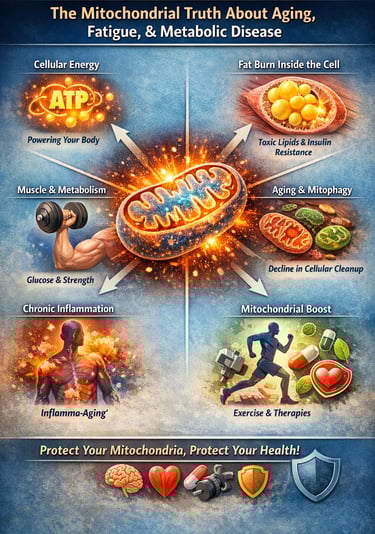
1️⃣ Your Cells Run on Energy — Not Just Calories
Mitochondria are the microscopic “power stations” inside your cells. They convert nutrients into ATP, the energy currency that powers muscle contraction, brain signaling, hormone regulation, and repair. When mitochondria function well, metabolism is flexible and resilient. When they falter, fatigue and metabolic dysfunction follow.
2️⃣ Fat Burning Must Happen Inside the Cell
Burning fat is not just about weight loss — it is about mitochondrial efficiency. When fat cannot be properly oxidized (a process called beta-oxidation), toxic lipid intermediates accumulate inside muscle cells. These compounds interfere with insulin signaling and promote inflammation. This is how metabolic disease often begins — silently, at the cellular level.
3️⃣ Muscle Is a Metabolic Organ
Skeletal muscle is your largest glucose-absorbing tissue. When fat accumulates inside muscle fibers and mitochondrial function declines, insulin resistance develops. Over time, this contributes to rising blood sugar, abdominal fat gain, and loss of metabolic flexibility.
4️⃣ Aging Slows Cellular “Cleanup”
Healthy cells regularly remove damaged mitochondria through a process called mitophagy. With aging, this quality-control system weakens. Dysfunctional mitochondria accumulate, producing excess oxidative stress and less usable energy. This contributes directly to sarcopenia (age-related muscle loss), frailty, and reduced endurance.
5️⃣ Inflammation Is an Energy Problem
When damaged mitochondria leak stress signals, the immune system becomes chronically activated. This low-grade inflammation — sometimes called “inflammaging” — is now linked to heart disease, type 2 diabetes, neurodegeneration, and physical decline.
6️⃣ The Encouraging Reality
Mitochondrial health is modifiable. Resistance training, aerobic exercise, metabolic balance, sleep optimization, and certain targeted therapies can stimulate mitochondrial biogenesis and improve efficiency. Unlike chronological aging, biological aging is dynamic.
Bottom Line:
Many chronic diseases are not simply the result of aging — they reflect an energy production crisis inside the cell. Protect the mitochondria, and you protect muscle, metabolism, brain function, and long-term vitality.
Summary: The 5 Pillars of Cellular Youth
To stay vital, your cells need to:
Burn fat cleanly (Beta-oxidation).
Keep muscles clear of "clogging" fats (IMCL management).
Stop energy leaks (ETC efficiency).
Take out the trash (Mitophagy).
Cool the flames of inflammation (Reducing inflammaging).
The Mitochondrial Mastery Checklist
1. Daily Metabolic Signaling (The "Basics")
Zone 2 Cardio (20–45 mins): Maintain a "conversational" pace to stimulate PGC-1α and improve mitochondrial fat-burning (Beta-oxidation).
Circadian Light Exposure: Get 5–10 minutes of morning sunlight to sync your mitochondrial "clocks" and optimize ATP production.
Hydration + Electrolytes: Ensure adequate Magnesium and Potassium intake to support the electrochemical gradient of the Electron Transport Chain (ETC).
Protein Pulse: Consume 25–40g of high-quality protein (leucine-rich) to overcome "anabolic resistance" and provide building blocks for mitochondrial enzymes.
2. Weekly Quality Control (Mitophagy & Stressors)
Resistance Training (2–3x/week): Activate the "Metabolic Sink" of your muscles to clear out stored fats (IMCLs) and trigger mitochondrial turnover.
Time-Restricted Feeding (14–16 hours): Allow at least one 16-hour fasting window weekly to trigger Mitophagy—the "cellular trash collection" that clears damaged mitochondria.
Cold/Heat Hormesis: One session of cold exposure (30s shower) or sauna (20 mins) to activate mitochondrial "uncoupling" proteins that improve metabolic flexibility.
Polyphenol Intake: Focus on "Mito-foods" like pomegranate (Urolithin A precursor), berries, and cruciferous vegetables to cool systemic inflammation.
3. Quarterly Monitoring (The "Bio-Metrics")
Check Fasting Insulin: A primary indicator of how well your "Metabolic Sink" is functioning. Aim for <5 μIU/mL.
Track HRV (Heart Rate Variability): Use your wearable to ensure your "RMSSD" is stable or improving—a sign of low internal oxidative stress.
Strength Benchmarks: Track your grip strength or leg press capacity; muscle function is a direct proxy for mitochondrial health in sarcopenia prevention.
The "Red Flag" Filter (What to Avoid)
Stagnant Fat: Avoid high-fat meals combined with a sedentary day (prevents "incomplete" fat burning/acylcarnitine buildup).
Late-Night Glucose Spikes: Avoid eating within 3 hours of sleep; high blood sugar during sleep "gums up" the mitochondrial repair process.
Chronic Over-Caffeination: Don't use caffeine to mask "bioenergetic failure" (cellular fatigue).
Clinical Pearl: Think of this checklist not as a "to-do" list, but as a maintenance schedule for your body’s power plants. If you don't clear the "smoke" (inflammation) and "take out the trash" (mitophagy), the power plants eventually fail, leading to the muscle loss and fatigue we call "aging."
Frequently Asked Questions (FAQs)
FAQ 1: What is beta-oxidation and why does its impairment matter?
Beta-oxidation is the mitochondrial process that breaks down long-chain fatty acids into acetyl-CoA, which then enters the citric acid cycle to generate ATP. When this process is impaired — due to enzyme dysregulation, substrate overload, or mitochondrial damage — cells cannot efficiently use fat for energy. Instead, fatty acid intermediates accumulate as acylcarnitines and lipid metabolites, triggering inflammation, oxidative stress, and insulin resistance. In a high-fat dietary environment, impaired beta-oxidation is a primary contributor to the metabolic dysfunction underlying type 2 diabetes and NAFLD (Aqeel et al., 2025).
FAQ 2: What are intramyocellular lipids (IMCLs) and how do they cause insulin resistance?
IMCLs are lipid droplets stored within muscle fibers — primarily in the form of ceramides, diacylglycerols (DAGs), and acylcarnitines. While trained endurance athletes accumulate IMCLs as a metabolic adaptation (the 'athlete's paradox'), in sedentary or obese individuals, IMCL accumulation reflects mitochondrial insufficiency. These lipid species directly inhibit insulin signaling by activating PKCθ and PP2A, which phosphorylate IRS-1 at inhibitory serine residues, blunting the insulin response. The inflammatory cytokines simultaneously generated create a compound barrier to glucose uptake in muscle — the body's largest glucose disposal organ (Razi et al., 2025).
FAQ 3: Can the electron transport chain (ETC) be targeted therapeutically?
Yes, and this is one of the most active areas of longevity science. ETC Complex I is the largest and most ROS-prone complex, and its decline with aging is both universal and consequential. Current interventions with clinical or preclinical evidence include CoQ10 supplementation (improves electron shuttling), NAD+ precursors such as NMN and NR (restore NADH/NAD+ ratios and re-energize Complex I), MitoQ (mitochondria-targeted antioxidant), and metformin (mild Complex I inhibition that paradoxically activates AMPK and mitochondrial quality control). Radovic and colleagues (2025) provide a thorough evidence-based ranking of these interventions by strength of evidence.
FAQ 4: What is mitophagy and why is its decline so damaging in aging?
Mitophagy is the cell's selective autophagy process for removing damaged or depolarized mitochondria before they can cause harm. The primary pathway — PINK1/Parkin — continuously monitors mitochondrial membrane potential and tags depolarized organelles for lysosomal degradation. With age, PINK1 expression falls, lysosomal acidification declines, and the entire mitophagy apparatus becomes sluggish. The result is progressive accumulation of dysfunctional mitochondria that generate excess ROS, release inflammatory mtDNA, and fail to meet cellular energy demands. Wang and colleagues (2026) identify mitophagy restoration — particularly through Urolithin A and spermidine — as one of the most clinically advanced longevity interventions currently available.
FAQ 5: How do mitochondria connect metabolic disease to brain aging and Alzheimer's disease?
Neurons are among the most metabolically demanding cells in the body, relying almost exclusively on mitochondrial ATP production for synaptic transmission, dendritic maintenance, and neuroplasticity. Age-related ETC decline reduces neuronal energy supply, impairing the energy-intensive processes of amyloid clearance (via the glymphatic system and proteasomal degradation). Simultaneously, dysfunctional mitochondria in neurons release DAMPs that activate neuroinflammatory pathways, promoting amyloid precursor protein (APP) misprocessing and tau hyperphosphorylation. Adlimoghaddam (2025) emphasizes that mitochondrial failure is not merely comorbid with Alzheimer's disease — it is mechanistically upstream of its pathological hallmarks.
FAQ 6: What is sarcopenia and why is mitochondrial dysfunction central to it?
Sarcopenia is the progressive, age-associated loss of skeletal muscle mass, strength, and function, affecting an estimated 10-27% of older adults globally and representing a major driver of frailty, falls, disability, and mortality. Mitochondrial dysfunction is now recognized as a core pathological mechanism in sarcopenia: declining ETC efficiency reduces ATP availability for protein synthesis; impaired mitophagy allows accumulation of ROS-generating mitochondria; reduced beta-oxidation capacity promotes IMCL accumulation; and mitochondria-driven inflammaging elevates catabolic cytokines (TNF-α, IL-6) that inhibit mTOR-mediated muscle protein synthesis. All five studies reviewed here directly implicate mechanisms relevant to sarcopenic muscle pathology.
FAQ 7: What lifestyle and pharmacological interventions have the strongest evidence for improving mitochondrial function?
Across the five studies reviewed, several interventions emerge with strong mechanistic rationale and accumulating clinical evidence. Exercise — particularly resistance training combined with endurance exercise — remains the single most potent mitochondrial biogenesis stimulant, upregulating PGC-1α and improving ETC efficiency. Caloric restriction and time-restricted eating enhance mitophagy through AMPK activation. Pharmacologically, metformin, NAD+ precursors (NMN/NR), CoQ10, Urolithin A (mitophagy inducer), and spermidine have demonstrated meaningful effects in human trials. Emerging gene and mitochondria transplantation therapies hold promise for more severe mitochondrial disorders.
Author’s Note
This article and accompanying infographic are intended for educational and informational purposes only. The content is based on peer-reviewed research published between 2025 and 2026, with a focus on mitochondrial biology, metabolic health, aging, and sarcopenia. While every effort has been made to present the science accurately and in a patient-friendly manner, this information does not replace professional medical advice, diagnosis, or treatment.
Readers are encouraged to discuss any health concerns, exercise programs, or dietary interventions with a qualified healthcare provider before making changes. Emerging therapies and supplements mentioned herein (e.g., NAD+ precursors, Urolithin A, CoQ10) have shown promise in research but may not be appropriate for all individuals, and their long-term effects are still under investigation.
The goal of this work is to provide accessible insights into the central role of mitochondrial health in overall well-being, illustrating how cellular energy production impacts metabolism, muscle function, brain health, and aging. By understanding these mechanisms, patients and clinicians alike can better appreciate the lifestyle, nutritional, and therapeutic strategies that support cellular vitality and long-term metabolic resilience.
Disclaimer: This article is for informational purposes only and does not constitute medical advice. Individual circumstances vary, and treatment decisions should always be made in consultation with qualified healthcare professionals.
Related Articles
Mitochondria: The Missing Link Between Fatigue, Weight Gain, and Brain Fog | DR T S DIDWAL
Vascular Calcification Is Not ‘Normal Aging’ — It’s Dysregulated Calcium Biology | DR T S DIDWAL
Sarcopenic Obesity: How to Lose Fat Safely Without Losing Muscle | DR T S DIDWAL
Why We Age: The Hidden Role of Chronic Inflammation in Accelerating Aging | DR T S DIDWAL
Vitamin D Deficiency and Sarcopenia: The Critical Connection | DR T S DIDWAL
References
Aqeel, A., Akram, A., Ali, M., Iqbal, M., Aslam, M., Rukhma, & Shah, F. I. (2025). Mechanistic insights into impaired β-oxidation and its role in mitochondrial dysfunction: A comprehensive review. Diabetes Research and Clinical Practice, 223, 112129. https://doi.org/10.1016/j.diabres.2025.112129
Razi, O., De Moraes, C., Zamani, N., Saeidi, A., Hadjicharalambous, M., Hackney, A. C., Del Coso, J., Laher, I., & Zouhal, H. (2025). From obesity to muscle insulin resistance: The mediating roles of intramyocellular lipids, inflammation, and oxidative stress. Diabetes/Metabolism Research and Reviews, 41(7), e70094. https://doi.org/10.1002/dmrr.70094
Radovic, M., Gartzke, L. P., Wink, S. E., van der Kleij, J. A., Politiek, F. A., & Krenning, G. (2025). Targeting the electron transport system for enhanced longevity. Biomolecules, 15(5), 614. https://doi.org/10.3390/biom15050614
Wang, Q., Sun, Y., Li, T. Y., et al. (2026). Mitophagy in the pathogenesis and management of disease. Cell Research, 36, 11-37. https://doi.org/10.1038/s41422-025-01203-7
Adlimoghaddam, A. (2025). Mitochondrial dysfunction in aging and age-related disorders. Aging and Disease, 16(5), 2495-2497. https://doi.org/10.14336/AD.2025.10731