Post-Meal Health: The Hidden 2-Hour Window That Predicts Heart Disease and Diabetes
Your fasting blood sugar might be normal, but what happens after you eat? Learn how "postprandial" glucose and triglycerides drive metabolic damage and how to fix it with simple habits.
DIABETES
Dr. T.S. Didwal, M.D.(Internal Medicine)
12/27/202515 min read
Most people think heart disease and diabetes are predicted by fasting blood tests taken once a year. Yet growing evidence suggests the most important damage happens after we eat—within a quiet 2-hour window when glucose and fats surge through the bloodstream.
During this post-meal period, blood vessels experience oxidative stress, inflammation, and impaired insulin signaling—often long before fasting glucose or HbA1c become abnormal (Wang et al., 2025; Reytor-González et al., 2025). Repeated several times daily, these hidden postprandial spikes gradually accelerate cardiovascular and metabolic disease.
Understanding what happens in the two hours after eating may be one of the most powerful—and overlooked—tools for preventing heart disease and diabetes.
Clinical pearls
1. The "2-Hour Window" is Your Metabolic Stress Test
Think of a meal as a "stress test" for your cells. While fasting labs show your body at rest, the 2-hour post-meal glucose measurement shows your body in action. Research now shows that if your glucose is above 155 mg/dL two hours after eating, your risk for future heart issues rises—even if your "A1c" or fasting results look perfect. It’s the difference between checking a car’s engine while it’s idling versus when it’s driving uphill.
2. Muscle is a "Sponge" for Sugar (But Only When Moving)
Your muscles are the primary destination for the sugar (glucose) in your blood. However, when you sit still after a meal, the "doors" to those muscle cells remain mostly shut, forcing your pancreas to overwork. By taking a simple 2-to-15-minute walk after lunch, you mechanically open those doors without needing extra insulin. This turns your muscles into a metabolic sponge, soaking up the "tsunami" of sugar before it can damage your arteries.
3. The "Sequence" of Your Bite Matters
The order in which you eat your food changes how your body processes it. Starting a meal with protein (like salmon or chicken) or fiber (like a salad) stimulates a gut hormone called GLP-1. This hormone slows down stomach emptying and primes the pancreas. If you save the refined carbohydrates (like bread or rice) for the end of the meal, you can reduce your post-meal glucose spike by up to 40% compared to eating the carbs first.
4. Triglycerides are the "Residual Risk" Hidden in Plain Sight
Even if your LDL (the "bad" cholesterol) is low, high levels of fat in the blood after a meal—called remnant lipoproteins—can still cause plaque buildup. These particles are smaller and more "sticky" than standard cholesterol. Using the TyG Index (a simple calculation using your fasting triglycerides and glucose) can help your doctor see if these "hidden" fats are putting you at risk, even when your standard cholesterol panel looks "normal."
5. Your Internal Clock Controls Your Insulin Sensitivity
Your body is biologically programmed to handle nutrients better in the morning than in the evening. As the sun goes down, your body naturally becomes more "insulin resistant" in preparation for sleep. This means a bowl of pasta at 9:00 PM causes a much higher and more damaging blood sugar spike than the exact same bowl of pasta at 12:00 PM. "Front-loading" your calories—eating a larger breakfast and a lighter, earlier dinner—aligns your habits with your DNA.
From Meal to Malfunction: Understanding Postprandial Cardiometabolic Health in 2025
Post-meal (postprandial) health plays a far more important role in predicting heart disease and diabetes than most people realize. After every meal, the body experiences a temporary surge in glucose, fats, and metabolic by-products. In healthy individuals, this surge is rapidly controlled. However, in many people—often years before abnormal fasting tests appear—these post-meal spikes linger and trigger harmful biological responses.
Recent research shows that 2-hour postprandial glucose levels predict diabetes, cardiovascular events, and even mortality more accurately than fasting glucose or HbA1c alone (Wang et al., 2025). During this critical window, impaired insulin signaling, delayed clearance of triglyceride-rich particles, oxidative stress, endothelial dysfunction, and inflammatory activation can quietly damage blood vessels (Reytor-González et al., 2025). Repeated several times daily, this “hidden stress” accelerates atherosclerosis and metabolic disease.
Importantly, these effects are modifiable. Mediterranean-style eating patterns, earlier meal timing, short walks after meals, and protein or fiber consumed before carbohydrates can significantly blunt post-meal glucose and lipid surges (Dimina & Mariotti, 2019). Simple tools such as the 2-hour glucose test or the triglyceride–glucose (TyG) index can uncover risk that fasting labs miss.
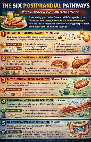
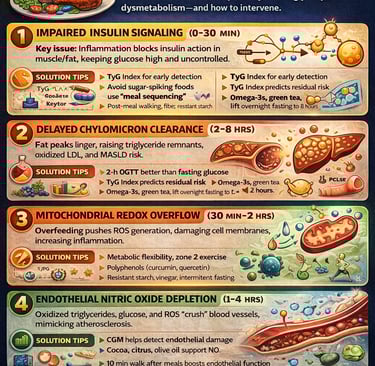
Modern eating patterns keep us in a near-constant postprandial state, with 4-10 eating occasions daily and minimal overnight fasting. This chronic metabolic stress cascades through six interconnected pathways, each a potential leverage point for intervention. Understanding these mechanisms isn't just academic—it's the key to preventing disease years before symptoms emerge.
1. Impaired Insulin Signaling (0-30 Minutes Post-Meal)
When you eat a mixed meal, your pancreas fires insulin. Normally, this hormone binds to receptors in muscle and fat cells, triggering a cascade that allows glucose transporters (GLUT4) to move to the cell surface and absorb glucose. This process relies on a precise molecular handshake: insulin receptor → IRS proteins → PI3K → Akt → GLUT4 trafficking.
In people with insulin resistance, this relay breaks down. Chronic inflammation serine-phosphorylates IRS proteins, preventing the normal tyrosine phosphorylation needed for PI3K recruitment. The result? Glucose stays in the bloodstream longer, fueling metabolic chaos downstream. Remarkably, this defect can occur while fasting insulin remains normal—a blindness in current diagnostics.
Key takeaway: Postprandial insulin signaling disruption happens faster than most standard tests can detect it.
2. Delayed Chylomicron Clearance (2-8 Hours Post-Meal)
After eating fat, your small intestine packages dietary triglycerides into vehicles called chylomicrons, which enter your bloodstream via lymph. Normally, an enzyme called lipoprotein lipase (LPL) rapidly breaks down these triglyceride-rich lipoproteins (TRLs), allowing tissues to uptake the released fatty acids. In a healthy person, this phase lasts 2-4 hours.
But insulin resistance inverts this process. It lowers LPL activity while raising inhibitors like ANGPTL3 and apolipoprotein C3 (APOC3). The result: postprandial triglyceride peaks can extend 8-12 hours, bathing your arteries in atherogenic remnant particles. These small, dense remnants penetrate the arterial wall, activate macrophages, and promote atherosclerosis even when LDL cholesterol is controlled.
For your liver, the impact is severe. High postprandial triglycerides dump excess fatty acids into hepatic circulation, driving the "dual-TRL hit" that fuels hepatic steatosis and MASLD progression.
Key takeaway: Postprandial 2-h glucose tolerance and postprandial triglyceride response jointly predict cardiovascular mortality and diabetes outcomes independent of fasting values.
3. Mitochondrial Redox Overflow (30 Minutes - 2 Hours Post-Meal)
A meal is an oxidative challenge. Within 60-180 minutes, mitochondria experience rapid substrate overflow—the cell's "redox overload." Electrons pile up at Complex I and III, generating a burst of reactive oxygen species (ROS). Normally, your cells neutralize this with antioxidant defenses (glutathione peroxidase, catalase, Nrf2-driven pathways).
In insulin-resistant phenotypes, this defense is blunted. The ROS burst is higher, longer, and unopposed. Superoxide quenches nitric oxide (NO) to form peroxynitrite, impairing vasodilation. Oxidized lipids and proteins accumulate. The damage window—typically 1-3 hours—becomes a gateway to chronic inflammatory signaling.
Metabolomics reveals this stress: acylcarnitine signatures spike during mixed meals, indicating mitochondrial redox pressure. In severe dysmetabolism, even modest meals trigger an ROS signature more commonly seen in acute illness.
Key takeaway: Repeated meals create cumulative redox stress; interventions targeting mitochondrial efficiency (resistant starch, exercise, polyphenols) can compress this window (Wu et al., 2025).
4. Endothelial Nitric Oxide Depletion (1-4 Hours Post-Meal)
The endothelium—the single-cell lining of your blood vessels—is the first interface to sense the postprandial surge. Under healthy conditions, it continuously releases nitric oxide, keeping blood vessels relaxed and pliable.
After a meal, glucose, remnant lipoproteins, and the ROS burst converge on the endothelium. NOX2/NOX4 enzymes are activated. Uncoupled eNOS generates superoxide instead of NO. The result: brachial-artery flow-mediated dilation (FMD)—a gold-standard marker of endothelial function—declines by approximately 1 percentage point at 2-4 hours post-meal.
This isn't a trivial number. Each 1% decline in FMD tracks with faster carotid intima-media thickness (IMT) progression and higher cardiovascular event risk. With three meals per day, you're damaging your endothelium three times daily.
Key takeaway: Postprandial endothelial dysfunction is reversible with Mediterranean dietary patterns and brief post-meal walking, yet completely missed by standard office testing.
5. NLRP3 Inflammasome Activation & Cytokine Release (2-6 Hours Post-Meal)
A phosphate-enriched meal doubles caspase-1 activity in monocytes, triggering the NLRP3 inflammasome—the cell's alarm bell for nutrient danger. Within hours, interleukin-1β (IL-1β) and interleukin-6 (IL-6) spike. Toll-like receptor 4 (TLR4), activated by saturated fats and remnant lipoproteins, upregulates adhesion molecules (VCAM-1, ICAM-1).
Older adults with cardiometabolic risk generate 40% higher IL-1β and CRP peaks than healthy peers. APOE ε4 carriers mount 2-fold greater postprandial CRP and endothelial-activation marker responses (Reytor-González et al., 2025). This innate-immune "memory" amplifies with age and repeated insults.
The practical consequence: chronic low-grade inflammation—"metaflammation"—accumulates from meal to meal. This feeds forward into insulin resistance, vascular stiffness, and β-cell exhaustion.
Key takeaway: Postprandial inflammation is highly modifiable; butyrate-producing microbes and post-meal activity can halve these peaks.
6. Microbiota-Bile-Acid-Hormone Integration (Hours to Days Post-Meal)
Your gut bacteria respond to every meal. Dysbiosis reshapes the bile-acid pool, modifying farnesoid X receptor (FXR) and Takeda G-protein-coupled receptor 5 (TGR5) signaling. Microbial short-chain fatty acids (SCFAs), especially butyrate, tune L-cell GLP-1 secretion. Indoles produced by tryptophan metabolism activate the aryl hydrocarbon receptor (AhR), influencing barrier function and immune tone.
In a landmark human study, lower butyrate output associated with sharper IL-6 and glycosylated acute-phase reactant peaks, an effect magnified by genetic variants in SCFA transporters (SLC5A8, FFAR2/3). This individual variation in microbiome-endocrine integration explains why identical meals elicit vastly different postprandial glucose and triglyceride responses between people.
Key takeaway: Microbiome composition is a modifiable biomarker of postprandial cardiometabolic stress; resistant starch and marine omega-3s reshape the microbiota within weeks.
Postprandial Glucose Tolerance and Postprandial 2-h Glucose Tolerance: The Hidden Predictor
A landmark 2025 study reveals that postprandial 2-h glucose tolerance is associated with diabetes diagnosis, diabetes mortality, and cardiovascular mortality—independent of fasting glucose or HbA1c (Wang et al., 2025). This is a paradigm shift.
Why is 2-hour glucose so predictive? At this timepoint, you're seeing the integrated response to insulin secretion, hepatic glucose output, and peripheral glucose uptake. A person with impaired postprandial glucose tolerance has already demonstrated metabolic incompetence to handle a meal.
Wang and colleagues showed that risk stratification by 2-h postprandial glucose outperforms traditional hemoglobin A1c cutoffs for identifying individuals destined for diabetes and cardiovascular events. Those with postprandial 2-h glucose > 155 mg/dL had substantially higher hazard ratios for both diabetes incidence and mortality.
The Postprandial Glucose Tolerance Test: A Clinical Tool Awaiting Integration
The oral glucose tolerance test (OGTT) has long been underutilized in clinical practice. Yet the 2-hour glucose during a 75-gram OGTT captures postprandial glucose tolerance more accurately than any fasting measure. In landmark cohort work, glycemic responses to identical test meals showed large between-person differences—yet within-person reproducibility was high, pointing to stable, heritable postprandial signatures (Wu et al., 2025).
This means two critical insights: (1) your body's meal response is predictable and personalized; (2) monitoring your own postprandial glucose response via continuous glucose monitoring (CGM) is far more informative than periodic fasting labs.
Clinical implication: Individuals with impaired postprandial glucose tolerance but normal fasting glucose are at elevated risk for both diabetes and cardiovascular disease. This is the "glucose blindness" that traditional screening misses.
Postprandial Cardiometabolic Biomarkers: Beyond Fasting Lipids and Glucose
The Triglyceride-Glucose Index (TyG): A Low-Cost, High-Impact Composite
The triglyceride-glucose (TyG) index—calculated as ln[fasting triglycerides (mg/dL) × fasting glucose (mg/dL) / 2]—is emerging as a cornerstone biomarker of postprandial cardiometabolic stress. Here's why it matters:
Dynamic signal: Higher fasting TyG predicts steeper 2-hour glucose and triglyceride rises on standardized meal tests, outperforming HOMA-IR
Cardiovascular outcomes: TyG ≥ 8.8 flags approximately 75% higher 5-year major adverse cardiovascular events in premature coronary disease
Stroke and progression: Elevated TyG associates with faster carotid intima-media thickness progression and higher incident ischemic stroke
Metabolic disease: In MASLD cohorts, TyG rivals triglyceride/HDL-C for detecting non-alcoholic fatty liver disease
The beauty of TyG is simplicity: no special assay needed, calculated from routine fasting labs, generalizable across populations.
Emerging Markers: Ceramides, GlycA, and Endothelial Vesicles
Recent metabolomic and inflammatory panels reveal nuanced information about postprandial metabolic stress:
Ceramides (especially C18:0/C24:0 ratio): Saturated ceramides rise after high-fat/refined-carb meals and predict insulin resistance conversion and cardiovascular events. Prospective data show ceramide C18:0/C24:0 + TyG as a potent composite for risk stratification.
GlycA (Glycoprotein Acetylation): This NMR-based marker integrates acute-phase glycoprotein glycosylation and outperforms high-sensitivity CRP for detecting low-grade systemic inflammation and predicting metabolic syndrome and coronary calcification.
Endothelial Extracellular Vesicles (ICAM-1+ EVs) and microRNAs (miR-126-3p, miR-210): These markers track endothelial injury and correlate with IMT progression, FMD decline, and mortality in severe cardiometabolic disease.
A pragmatic tiered workflow for risk assessment:
Screen with TyG and triglyceride/HDL-C ratio
Stratify intermediate-risk patients with GlycA and endothelial-vesicle counts
Personalize high-risk cases with ceramide panels and microbiome-guided interventions
Evidence-Based Interventions for Postprandial Cardiometabolic Health
Mediterranean-Style Eating: The Gold Standard
The Mediterranean dietary pattern—extra-virgin olive oil, vegetables, legumes, whole grains, fish, and modest red wine—consistently blunts postprandial turbulence (Reytor-González et al., 2025). In a randomized crossover, a Mediterranean-type meal preserved endothelial function and attenuated triglyceride excursions versus a high-saturated-fat comparator. Importantly, fine-tuning within the Mediterranean framework amplifies benefits:
Carbohydrate quality: Replacing refined starches with low-glycemic-index pulses during an isocaloric Mediterranean day blunted postprandial glucose and insulin excursions during an 8-hour mixed-meal tolerance test
Fat quality: Swapping saturated fat for monounsaturated fat (EVOO, nuts) shifted the postprandial metabolomic profile toward lower acylcarnitines and higher antioxidant signals
Exercise synergy: Adding 150 min/week of brisk walking to Mediterranean eating improved the lipoprotein subclass profile, lowering fasting triglycerides and small dense LDL
Chrononutrition and Meal Timing: Aligning Meals with Your Body Clock
Timing matters as much as content. Front-loading energy in the morning and tapering evening carbohydrates blunts both glycemic and lipemic excursions:
Skipping breakfast increases lunchtime and dinnertime glycemic excursions in type 2 diabetes, accompanied by higher glucagon and lower iGLP-1
Shifting the main meal earlier—an early dinner at 18:00 versus 21:00—lowers 24-hour mean glucose and increases next-morning fat oxidation at identical calorie intake
Early time-restricted eating (eTRE) (e.g., 08:00-14:00 window) reduced 24-hour mean glucose and glycemic variability without weight loss
Within-meal sequencing also matters: A 15-gram whey protein preload taken 10 minutes before breakfast reduced the 0-240 minute glucose iAUC
and increased insulin/GLP-1 in type 2 diabetes. Across controlled-feeding studies, starting meals with protein or fat ("protein-first/fat-first") consistently lowers early postprandial glucose versus carbohydrate-first sequences.
Physical Activity "Snacks": Turning Muscle Into a Second Pancreas
Even brief muscle contractions stimulate GLUT4 translocation and LPL activation. Breaking up sitting with 2-5 minute bouts of light walking every 20-30 minutes lowers both postprandial glucose and insulin versus uninterrupted sitting. A meta-analysis of approximately 22 randomized trials confirms this effect across mixed-risk adults.
Post-meal walking is particularly potent: a single 15-minute walk after lunch more effectively controls postprandial glycemia than a continuous 45-minute morning walk at equivalent intensity.
Dietary Bioactives: Rapid-Response Molecules
Plant-derived polyphenols can blunt oxidative and metabolic surges within minutes (Dimina & Mariotti, 2019):
Catechins + chlorogenic acids: Co-ingestion of combined polyphenols (150-300 mg) produced graded reduction in early postprandial glycemia in healthy men
Anthocyanin-rich red raspberries: Test meals containing 0, 125, or 250 grams of red raspberries produced dose-dependent improvements in postprandial glucose and insulin dynamics
Curcumin (longer-term): Meta-analysis shows curcumin supplementation (80-1,000 mg/day for ≥4 weeks) lowers fasting glucose, CRP, and overall glycemic indices
For acute control, a 150-300 mg catechin/chlorogenic acid blend taken before carbohydrate-rich meals helps dampen early glycemic excursions (0-2 hours).
Macronutrient Manipulation: Quality Over Quantity
Swap refined carbohydrates for monounsaturated/polyunsaturated fat: Replacing 10% of carbohydrates with unsaturated fat reduces postprandial glucose AUC by approximately 12%
Protein preload: Approximately 20 grams of whey taken 15 minutes pre-meal lowers glucose iAUC by approximately 12%
Resistant starch: RS4 (phosphorylated wheat) acutely lowers incremental insulin iAUC and attenuates the second-meal glucose peak; practical intake: 15-30 g/day
Study Summaries: Key Takeaways from 2025 Research
Reytor-González et al. (2025) - Frontiers in Cardiovascular Medicine
This comprehensive review synthesized human studies from 2020-2025, identifying six interconnected pathways of postprandial dysmetabolism. The authors emphasize that dynamic, after-meal markers (e.g., triglyceride-glucose index, ceramides, GlycA) outperform fasting measures for identifying risk. Mediterranean-style meals, early energy distribution, pre-meal protein, and post-meal walking were endorsed as practical strategies to shorten the "damage window" within hours after eating.
Key takeaway: A tiered workflow—screen, stratify, personalize—reframes prevention around after-meal physiology, with relevance to resource-limited settings.
Wang et al. (2025) - Scientific Reports
This landmark study of postprandial 2-h glucose tolerance in over 10,000 adults revealed that elevated 2-hour glucose during an OGTT predicts diabetes diagnosis, diabetes mortality, and cardiovascular mortality independent of fasting glucose or HbA1c. Those with postprandial 2-h glucose > 155 mg/dL faced substantially higher hazard ratios.
Key takeaway: Postprandial glucose tolerance testing captures metabolic dysfunction earlier than fasting metrics and warrants wider clinical integration.
Dimina & Mariotti (2019) - Nutrients
While foundational, this review articulated the mechanisms by which postprandial appearance of cardiometabolic risk features occurs acutely and is preventable by nutrient and dietary-substance interventions. The authors detailed how postprandial hyperglycemia, hypertriglyceridemia, and hyperinsulinemia trigger oxidative stress, inflammation, and endothelial dysfunction within hours.
Key takeaway: Postprandial dysmetabolism is not merely a fasting-state problem; acute meal-induced stresses are modifiable biomarkers of prevention opportunity.
Tudorache et al. (2025) - European Atherosclerosis Journal
This review identified postprandial markers (TyG, ceramides, endothelial-activation markers) as potentially useful for developing new cardiovascular therapies. Authors highlighted that standard fasting assessments miss residual atherosclerotic risk in controlled LDL-C states, emphasizing that postprandial TRL dynamics drive much of this "residual" risk.
Key takeaway: Novel CVD prevention strategies should target postprandial lipoprotein metabolism, not only fasting lipids.
Wu et al. (2025) - Nature Medicine
This translational study revealed individual variations in glycemic responses to carbohydrates reflect underlying differences in metabolic physiology (pancreatic insulin secretion capacity, insulin sensitivity, hepatic glucose output). Multiomics and machine-learning models explained approximately 40% of between-person variance in postprandial glucose iAUC, far exceeding glucose-only predictors.
Key takeaway: Postprandial glucose phenotyping—informed by metabolomics and microbiome data—enables truly personalized dietary recommendations, outperforming one-size-fits-all guidelines.
FAQs: Your Postprandial Cardiometabolic Health Questions Answered
Q: What's the difference between fasting glucose and postprandial glucose?
A: Fasting glucose reflects liver glucose output during overnight fasting and may be normal even in people with severe insulin resistance. Postprandial glucose, measured 2 hours after a meal, reflects your body's ability to handle food—a much better predictor of diabetes risk and cardiovascular events.
Q: Can I lower my postprandial glucose without medication?
A: Absolutely. Mediterranean eating, post-meal walking (even 2-5 minutes), pre-meal protein, and resistant starch collectively flatten glucose and triglyceride peaks as effectively as some diabetes medications. Combine these tactics for maximal effect.
Q: How often should I test my postprandial glucose?
A: If you have risk factors (family history, obesity, prediabetes), one baseline 2-hour glucose tolerance test (75-gram OGTT) is informative. Continuous glucose monitoring (CGM) or home glucose monitoring after meals can further personalize dietary strategies.
Q: Is the triglyceride-glucose index better than standard lipid panels?
A: TyG is superior for predicting cardiovascular events and metabolic dysfunction compared to LDL-C alone, especially in people with obesity or metabolic syndrome. It costs nothing extra (calculated from fasting labs) and should be standard in clinical practice.
Q: Which dietary pattern is best for postprandial health?
A: Mediterranean eating is gold-standard, but the key is early meal timing (breakfast > dinner), low-glycemic-index carbs, unsaturated fats, adequate protein, and post-meal movement. Personalization based on your microbiome composition and glucose phenotype enhances results further.
Q: Can I reverse postprandial dysmetabolism?
A: Yes. Within weeks of adopting Mediterranean eating + exercise, endothelial function improves, postprandial glucose and triglyceride peaks flatten, and inflammatory markers decline. The damage window—1-3 hours post-meal—becomes progressively shorter and less severe.
Key Takeaways: What You Must Know About Postprandial Cardiometabolic Health
Fasting labs lie. Normal fasting glucose masks dangerous postprandial glucose surges that predict cardiovascular disease and diabetes. A 2-hour glucose during an OGTT or home monitoring after meals reveals your true metabolic risk.
The damage window is real and modifiable. Within 1-6 hours after eating, your endothelium is being injured, your mitochondria are generating oxidative stress, and your immune system is being triggered. This window is where prevention happens.
TyG is your new metabolic vital sign. Calculate this simple index from fasting labs (triglycerides × glucose ÷ 2, then natural log). Elevated TyG predicts cardiovascular events better than LDL-C and costs nothing.
Timing beats total calories. Front-loading breakfast and tapering evening meals flattens glucose/triglyceride peaks independent of total energy intake. Meal timing is a free, powerful lever.
Post-meal movement is medicine. A 2-5 minute walk after lunch more effectively controls postprandial glycemia than a morning gym session. Micro-exercise breaks are practical and profound.
Your microbiome predicts your postprandial response. Butyrate-producing bacteria explain significant variance in individual postprandial glucose tolerance. Resistant starch, marine omega-3s, and fiber reshape the microbiota within weeks and flatten glucose peaks.
Prevention is personalized. Identical meals trigger vastly different glucose and triglyceride responses between people. Continuous glucose monitoring + simple dietary adjustments enable truly individualized prevention before disease emerges.
Call to Action: Take Control of Your Postprandial Cardiometabolic Health Today
If you have risk factors for cardiometabolic disease—family history, obesity, prediabetes, or cardiovascular disease—don't wait for traditional screening to fail. Here's a practical roadmap:
This month:
Ask your doctor for a 2-hour glucose tolerance test or home glucose meter to assess your postprandial glucose response
Calculate your TyG index from latest fasting labs; if ≥ 8.5, prioritize the changes below
Next 4 weeks:
Shift to Mediterranean eating (EVOO, vegetables, legumes, whole grains, fish)
Front-load breakfast; taper evening meals
Add a 15-minute walk after lunch
Pre-meal protein (15 g whey) with carbohydrate-rich meals
By month 3:
Repeat postprandial glucose testing; expect flattened peaks
Consider a stool microbiome test (optional but informative)
Add resistant starch (15-30 g daily) to amplify benefits
The evidence is clear: postprandial cardiometabolic stress is the hidden driver of chronic disease in 2025. By understanding the six molecular pathways, measuring the right biomarkers, and acting on evidence-based interventions, you can prevent or reverse metabolic dysfunction years before symptoms emerge.
Your next meal is not just fuel—it's an opportunity to either build health or accelerate disease. The choice, informed by science, is yours.
Disclaimer: This article is for informational purposes only and does not constitute medical advice. Individual circumstances vary, and treatment decisions should always be made in consultation with qualified healthcare professionals.
Related Articles
The Metabolic Triad: Why Diabetes, Obesity & CVD Are One Epidemic | DR T S DIDWAL
What’s New in the 2025 Blood Pressure Guidelines? A Complete Scientific Breakdown | DR T S DIDWAL
Manage Diabetes Naturally: How Beta-Glucans Control Blood Sugar | DR T S DIDWAL
Exercise as Metabolic Medicine: Latest Research on Glucose and Heart Health| DR T S DIDWAL
References
Dimina, L., & Mariotti, F. (2019). The postprandial appearance of features of cardiometabolic risk: Acute induction and prevention by nutrients and other dietary substances. Nutrients, 11(9), 1963. https://doi.org/10.3390/nu11091963
Reytor-González, C., Cevallos-Fernández, E., Jácome, B., & Simancas-Racines, D. (2025). From meal to malfunction: Exploring molecular pathways, biomarkers and interventions in postprandial cardiometabolic health. Frontiers in Cardiovascular Medicine, 12, Article 1655889. https://doi.org/10.3389/fcvm.2025.1655889
Tudorache, I. F., Barbarossa, D., Daraban, B. S., & Rizzo, R. (2025). Postprandial implications in cardiovascular disease and potential markers to develop new therapies: Postprandial state and novel CVD markers. European Atherosclerosis Journal, 4(2), Article 104. https://doi.org/10.56095/eaj.v4i2.104
Wang, Y., Fang, Y., Yang, G., et al. (2025). Postprandial 2-h glucose tolerance is associated with diabetes diagnosis, diabetes mortality, and cardiovascular mortality. Scientific Reports, 15, Article 43853. https://doi.org/10.1038/s41598-025-28849-y
Wu, Y., Ehlert, B., Metwally, A. A., et al. (2025). Individual variations in glycemic responses to carbohydrates and underlying metabolic physiology. Nature Medicine, 31, 2232–2243. https://doi.org/10.1038/s41591-025-03719-2