Ferroptosis Explained: The Iron-Driven Cell Death Linked to Cancer, Aging & Alzheimer's
Ferroptosis is an iron-driven form of cell death linked to cancer, Alzheimer's disease, aging, obesity, and inflammation. Discover how iron, diet, and antioxidants influence this emerging pathway.
AGING
Dr. T.S. Didwal, M.D.(Internal Medicine)
6/7/202620 min read
Ferroptosis is a form of iron-dependent cell death caused by the accumulation of lipid peroxides in cell membranes. Emerging research links ferroptosis to cancer, Alzheimer's disease, Parkinson's disease, aging, immunity, and obesity, making it one of the most important new targets in precision medicine and longevity science.
Key Takeaways: What Ferroptosis Means for Your Health
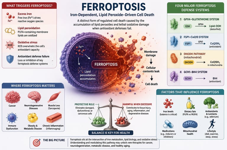
Ferroptosis is a unique form of regulated cell death driven by iron and lipid peroxidation. Unlike apoptosis ("programmed cell death"), ferroptosis occurs when iron-dependent oxidative damage overwhelms a cell's antioxidant defenses, causing the cell membrane to fail.
Iron, polyunsaturated fats (PUFAs), and antioxidant defenses determine whether ferroptosis occurs. Excess free iron and oxidized membrane fats promote ferroptosis, while protective systems such as GPX4, glutathione, CoQ10, and BH4 help prevent it.
Ferroptosis is neither inherently good nor bad. In healthy tissues, it helps eliminate damaged, dysfunctional, or potentially cancerous cells. However, excessive ferroptosis may contribute to tissue injury, chronic inflammation, and age-related disease.
Many cancers appear highly vulnerable to ferroptosis. Researchers are actively developing therapies that trigger ferroptosis in treatment-resistant tumors, making it one of the most promising emerging areas in cancer biology.
Several ferroptosis-defense systems protect cells from oxidative damage. These include the GPX4-glutathione pathway, the FSP1-CoQ10 system, the mitochondrial DHODH pathway, and the GCH1-BH4 pathway. Together, they form a multilayered defense network against uncontrolled cell death.
Ferroptosis may play an important role in aging and longevity. Excessive iron accumulation, declining antioxidant capacity, and increased oxidative stress with age may increase susceptibility to ferroptosis, potentially contributing to biolgical aging.
Growing evidence links ferroptosis to neurodegenerative diseases. Abnormal iron accumulation and ferroptotic damage have been observed in Alzheimer's disease and Parkinson's disease, suggesting that ferroptosis may contribute to progressive neuronal loss.
Ferroptosis may also contribute to age-related muscle loss (sarcopenia). Increased lipid peroxidation, mitochondrial dysfunction, and iron dysregulation in aging muscle may accelerate muscle decline through ferroptotic mechanisms.
Ferroptosis sits at the intersection of iron metabolism, oxidative stress, immunity, aging, neurodegeneration, and cancer. The goal is not to maximize or eliminate ferroptosis, but to maintain the delicate balance that allows damaged cells to be removed while protecting healthy tissues. As research advances, ferroptosis may become one of the most important therapeutic targets in oncology, longevity science, and metabolic medicine.
For decades, scientists believed they understood the major ways cells die. Then researchers discovered something entirely different: a process called ferroptosis, where cells are destroyed by iron-driven oxidative damage.
At first, ferroptosis seemed like a niche laboratory finding. Today, it is one of the hottest topics in biomedical research. Studies now link ferroptosis to cancer, Alzheimer's disease, Parkinson's disease, age-related muscle loss, chronic inflammation, obesity, and metabolic dysfunction. Researchers are even developing therapies designed to trigger ferroptosis in cancer cells while preventing it in healthy tissues.
What makes ferroptosis especially fascinating is that it sits at the crossroads of nutrition, iron metabolism, oxidative stress, immunity, and aging. Nutrients such as selenium, dietary fats, and antioxidants can influence the cellular pathways that regulate ferroptosis, although much remains to be learned.
As scientists unravel this complex process, ferroptosis is emerging as a potential key to understanding both disease prevention and healthy longevity.
Ferroptosis—the process of cells "rusting" from the inside out—may help explain cancer, Alzheimer's disease, obesity, aging, and why iron metabolism is so important for long-term health.
What Is Ferroptosis? The “Iron Death” Pathway Explained
Ferroptosis was officially named in 2012 by Dr Brent Stockwell’s lab at Columbia University. The name combines ferro, Latin for iron, and ptosis, Greek for falling.
Here’s the 10-second version: Too much free iron inside a cell causes fats in the cell membrane to oxidize. If the cell can’t stop the chain reaction, the membrane ruptures and the cell dies.
This is different from apoptosis, your body’s normal “programmed” cell death. Apoptosis is clean and tidy. Ferroptosis is like a grease fire — messy, inflammatory, and driven by metabolism.
Why evolution kept it: Ferroptosis likely evolved as a defense against cancer and infection. A cell infected with a virus or with damaged DNA can be forced to undergo ferroptosis before it becomes a bigger problem.
The 3 Core Mechanisms: Iron, Lipids, and GPX4
Think of ferroptosis as a 3-legged stool. You need all 3 for it to happen:
1. Iron (The Catalyst)
Role: Drives the chemical reaction behind ferroptosis.
Key Detail: Free ferrous iron triggers the Fenton reaction, which generates highly reactive hydroxyl radicals. These radicals launch the initial attack on cellular fats.
2. Polyunsaturated Fatty Acids (The Fuel)
Role: Acts as the "fuel" that catches fire.
Key Detail: Membrane lipids containing polyunsaturated fatty acids (PUFAs)—such as arachidonic acid—are uniquely vulnerable to oxidative damage because of their chemical structure.
3. Low GPX4 Activity (The Failed Defense)
Role: The breakdown of the cell's antioxidant shield.
Key Detail: Glutathione peroxidase 4 (GPX4) is the primary enzyme responsible for "putting out" lipid lipid-peroxidation fires. When GPX4 activity drops or is inhibited, the cell loses its defense, allowing lethal lipid damage to accumulate and trigger cell death.
The Four Major Ferroptosis Defense Systems
Cells possess multiple defense mechanisms that prevent uncontrolled ferroptosis by limiting lipid peroxidation and oxidative damage. While GPX4 was once considered the primary protector against ferroptosis, researchers now recognize several complementary systems that work together to maintain cellular survival.
1. GPX4–Glutathione System
The GPX4–glutathione pathway is considered the classical and most extensively studied ferroptosis defense mechanism. Glutathione peroxidase 4 (GPX4) uses glutathione (GSH) to neutralize toxic lipid peroxides before they can damage cell membranes. When glutathione levels fall or GPX4 activity is inhibited, lipid peroxides accumulate rapidly, triggering ferroptosis. This pathway depends heavily on adequate intake of sulfur-containing amino acids and the trace mineral selenium, which is required for GPX4 synthesis.
2. FSP1–CoQ10 System
Discovered in 2019, Ferroptosis Suppressor Protein 1 (FSP1) provides an alternative defense mechanism independent of GPX4. FSP1 regenerates reduced coenzyme Q10 (CoQ10), a potent lipid-soluble antioxidant located within cell membranes. Reduced CoQ10 acts as a "radical trap," intercepting lipid-free radicals before they initiate destructive chain reactions. This pathway helps explain why some cells remain resistant to ferroptosis even when GPX4 activity is compromised.
3. DHODH Pathway
Dihydroorotate dehydrogenase (DHODH) is a mitochondrial enzyme that protects against ferroptosis specifically within mitochondria. DHODH supports the regeneration of CoQ10 in the inner mitochondrial membrane, helping neutralize oxidative stress where reactive oxygen species are frequently generated. This discovery highlighted the importance of mitochondrial-specific ferroptosis defenses and revealed a crucial link between mitochondrial health and ferroptosis regulation.
4. GCH1–BH4 System
The GTP cyclohydrolase 1 (GCH1)–tetrahydrobiopterin (BH4) pathway represents another independent ferroptosis suppression system. BH4 functions as a powerful endogenous antioxidant that directly prevents lipid peroxidation and helps maintain membrane integrity. Beyond its role in neurotransmitter synthesis and nitric oxide production, BH4 can protect cells from ferroptotic death even when GPX4 activity is reduced, providing an additional layer of cellular protection.
Key Takeaway
Rather than relying on a single defense mechanism, cells use multiple overlapping systems—including GPX4–glutathione, FSP1–CoQ10, DHODH, and GCH1–BH4—to prevent excessive ferroptosis. Dysfunction in one or more of these pathways may contribute to cancer, neurodegeneration, aging, and chronic inflammatory diseases, making them promising therapeutic targets for future precision medicine
Ferroptosis and Disease: What the Research Shows
Cancer: Using Ferroptosis to Kill Tumors
Many aggressive cancers, like triple-negative breast cancer and pancreatic cancer, are resistant to apoptosis. But they have an Achilles’ heel: they’re addicted to iron and vulnerable to ferroptosis.
A 2020 study in Nature found that the drug erastin triggers ferroptosis by blocking cystine uptake, starving cells of glutathione. Newer research is testing iron nanoparticles and GPX4 inhibitors as cancer therapies.
Practical takeaway: You cannot “treat cancer with diet” alone. But understanding ferroptosis helps explain why some metabolic therapies are being studied in oncology. Always work with your oncologist.
Autoimmune Diseases: When Too Much Ferroptosis Hurts
If ferroptosis kills cancer, is more always better? No.
In diseases like lupus, rheumatoid arthritis, and multiple sclerosis, researchers have found excess ferroptosis in healthy tissues. A 2023 bibliometric analysis of 2018-2023 papers showed ferroptosis research in autoimmunity grew 400% in 5 years. The theory: immune cells and target tissues die from lipid peroxidation, worsening inflammation.
This is why indiscriminately taking high-dose iron or “ferroptosis-promoting” supplements is dangerous if you have autoimmunity.
Immune Function: How Diet Alters T Cell Ferroptosis
Your immune system walks a tightrope. A 2024 study in Cell Metabolism showed that T cells need controlled lipid peroxidation to function, but too much pushes them into ferroptosis.
Diets high in certain PUFAs, like arachidonic acid from animal fats, made T cells more prone to ferroptosis in mice. Diets rich in monounsaturated fats like olive oil were protective. Human data is still early, but this may explain some links between diet and immunity.
A 2026 study published in Nature further strengthened this concept by demonstrating that dietary lipid composition directly influences T-cell susceptibility to ferroptosis. Diets rich in polyunsaturated fats increased ferroptotic vulnerability, whereas monounsaturated fats appeared protective. These findings suggest that dietary fat quality may play an important role in regulating immune resilience through ferroptosis-related mechanisms.
Ferroptosis and Metabolic Health
Emerging evidence suggests that ferroptosis may also influence obesity and energy metabolism. A 2025 Cell Metabolism study found that controlled ferroptotic signaling from adipocytes helped improve metabolic flexibility and limit excessive fat accumulation in experimental models. These findings indicate that ferroptosis may function not only as a cell-death mechanism but also as a regulator of whole-body metabolic health.
Emerging Research: Ferroptosis, Immunity, and Metabolic Health
Dietary Fats, T-Cell Ferroptosis, and Immune Function
A landmark 2026 study by Wang and colleagues published in Nature demonstrated that dietary fat composition can directly influence immune-cell susceptibility to ferroptosis. The researchers found that diets enriched in polyunsaturated fatty acids (PUFAs) increased lipid peroxidation in T cells, making them more vulnerable to ferroptotic death. In contrast, monounsaturated fats appeared to protect T cells by reducing membrane susceptibility to oxidative damage. These findings suggest that dietary fat quality—not simply fat quantity—may influence immune resilience through ferroptosis-related mechanisms. Although the study provides important mechanistic insights, clinical studies are still needed to determine whether these effects translate into meaningful health outcomes in humans.
Ferroptosis and Obesity
A 2025 study published in Cell Metabolism reported that ferroptotic signaling originating from adipocytes (fat cells) may help counteract obesity and metabolic dysfunction. The researchers observed that controlled ferroptosis within dysfunctional adipose tissue triggered signaling pathways that enhanced energy expenditure, improved metabolic flexibility, and reduced excessive fat accumulation in experimental models. These findings challenge the traditional view that all cell death is harmful and suggest that carefully regulated ferroptosis may serve as a protective mechanism against obesity. While promising, these results are currently based primarily on preclinical research and should not be interpreted as evidence that inducing ferroptosis is a safe weight-loss strategy in humans.
Key Takeaway
Together, these studies highlight the emerging role of ferroptosis as a metabolic regulator that influences not only cancer and aging but also immune function, dietary responses, and obesity. Researchers increasingly view ferroptosis as a critical intersection between nutrition, metabolism, and chronic disease, making it one of the most rapidly evolving fields in biomedical science.
How to Modulate Ferroptosis Naturally: Diet & Lifestyle
Important: Modulate doesn’t mean “biohack.” For most people, the goal is balance — not maximum or minimum ferroptosis. Consult your doctor before changing iron intake or taking new supplements, especially with cancer, anemia, or autoimmune conditions.
Ferroptosis and Healthy Aging
Ferroptosis has emerged as a key biological process linking aging, neurodegenerative diseases, muscle loss, and chronic inflammation. While controlled ferroptosis helps eliminate damaged cells, excessive ferroptosis may contribute to tissue degeneration and age-related disease.
As we age, iron tends to accumulate in tissues while antioxidant defenses such as glutathione and GPX4 decline. This creates an environment that favors lipid peroxidation and ferroptotic cell death. Researchers increasingly view ferroptosis as a potential driver of biological aging and age-related dysfunction.
Alzheimer's Disease
Evidence suggests that abnormal iron accumulation occurs in brain regions affected by Alzheimer's disease. Excess iron promotes oxidative damage, lipid peroxidation, and neuronal ferroptosis, potentially accelerating cognitive decline. Postmortem studies have identified increased iron levels and markers of ferroptosis in Alzheimer's brains, making ferroptosis inhibition a promising therapeutic target.
Parkinson's Disease
The substantia nigra, the brain region most affected in Parkinson's disease, contains high concentrations of iron. Ferroptosis may contribute to the progressive loss of dopamine-producing neurons through iron-driven oxidative stress and mitochondrial damage. Experimental studies show that ferroptosis inhibitors can reduce neuronal death and improve outcomes in animal models of Parkinson's disease.
Sarcopenia (Age-Related Muscle Loss)
Emerging evidence indicates that ferroptosis may play a role in sarcopenia. Aging skeletal muscle often exhibits increased iron accumulation, impaired mitochondrial function, and elevated lipid peroxidation. Excessive ferroptosis may contribute to the loss of muscle fibers and reduced muscle strength observed in older adults.
Mitochondrial Dysfunction
Mitochondria are both sources and targets of oxidative stress. During aging, dysfunctional mitochondria generate increased reactive oxygen species (ROS), which can trigger lipid peroxidation and ferroptosis. Conversely, ferroptosis can further damage mitochondria, creating a vicious cycle that accelerates cellular aging and tissue dysfunction.
Inflammaging
Aging is characterized by chronic, low-grade inflammation, often referred to as "inflammaging." Ferroptotic cells release inflammatory molecules and oxidized lipids that can stimulate immune responses. Excessive ferroptosis may therefore contribute to the persistent inflammation associated with cardiovascular disease, neurodegeneration, frailty, and other age-related conditions.
Healthy aging likely requires a delicate balance: enough ferroptosis to remove damaged or potentially cancerous cells, but not so much that it drives neurodegeneration, muscle loss, mitochondrial dysfunction, and chronic inflammation. Maintaining iron homeostasis, supporting antioxidant defenses, regular physical activity, and consuming a nutrient-dense diet may help preserve this balance and promote healthy longevity.
Evidence Strength: What the Science Actually Shows
Ferroptosis can kill cancer cells — Strong Preclinical Evidence
Extensive laboratory and animal research demonstrates that inducing ferroptosis can effectively destroy many cancer cell types, including tumors that resist conventional therapies. This is one of the most well-established findings in ferroptosis research.Ferroptosis-targeting drugs may become future cancer treatments — Emerging Clinical Evidence
Several ferroptosis-inducing drugs and therapeutic strategies are being evaluated in early human studies. While results are promising, clinical evidence remains limited and no ferroptosis-specific therapy has yet become standard medical practice.Diet directly controls ferroptosis in humans — Weak Evidence
Although nutrients such as iron, selenium, antioxidants, and dietary fats influence ferroptosis-related pathways, there is currently little direct human evidence showing that specific diets can reliably increase or decrease ferroptosis in clinical settings.Olive oil may reduce susceptibility to ferroptosis — Moderate Mechanistic Evidence
Experimental studies suggest that monounsaturated fats, particularly those found in olive oil, are less prone to lipid peroxidation than many polyunsaturated fats. This may help make cell membranes more resistant to ferroptotic damage, although direct human evidence is limited.Selenium supports GPX4, the body's primary ferroptosis-defence enzyme — Strong Evidence
Selenium is an essential component of GPX4, a key enzyme that protects cells from ferroptosis. Numerous cellular, animal, and human studies confirm that adequate selenium status is critical for optimal GPX4 activity and antioxidant defense.
Current evidence strongly supports the biological importance of ferroptosis in cancer and the central role of selenium-dependent GPX4 defenses. However, claims that specific foods, supplements, or dietary patterns can precisely control ferroptosis in humans remain largely unproven and require further clinical investigation.
How to Modulate Ferroptosis Naturally: Diet & Lifestyle
Important: Modulate doesn’t mean “biohack.” For most people, the goal is balance — not maximum or minimum ferroptosis. Consult your doctor before changing iron intake or taking new supplements, especially with cancer, anemia, or autoimmune conditions.
Foods That Promote Ferroptosis
These increase iron availability or PUFA oxidation:
Red meat, organ meats: Heme iron is highly bioavailable
High-PUFA seed oils: Corn, soybean, sunflower oil oxidize easily when heated
Alcohol: Increases iron absorption and depletes glutathione
Foods & Nutrients That Block It
These boost antioxidant defenses or chelate iron:
Vitamin E & selenium: Direct cofactors for GPX4
Polyphenols: Green tea EGCG, curcumin, and quercetin are iron chelators
Monounsaturated fats: Olive oil and avocados displace oxidation-prone PUFAs in membranes
NAC & glycine: Support glutathione synthesis, fueling GPX4
Exercise, Fasting, and Other Modulators
Zone 2 cardio: Mildly increases lipid peroxidation short-term, but upregulates antioxidant defenses long-term
Intermittent fasting: May increase ferroptosis sensitivity in cancer cells while protecting healthy cells, per 2023 mouse data. Human trials ongoing
.
Supplements and Ferroptosis: What Works, What’s Hype
Promotes Ferroptosis (Drives Cell Death)
Iron bisglycinate
Evidence Level: Human RCTs (Randomized Controlled Trials) for anemia.
Mechanism: Actively promotes ferroptosis by supplying the raw $Fe^{2+}$ required to drive lipid peroxidation.
⚠️ Safety Note: Avoid unless you have a diagnosed deficiency. Excess iron significantly drives systemic oxidative stress.
Artemisinin
Evidence Level: Cancer cell lines (in vitro).
Mechanism: Promotes ferroptosis, which is why it is heavily researched in oncology to kill cancer cells.
⚠️ Safety Note: This is a potent drug, not a standard supplement. Do not self-prescribe.
Blocks Ferroptosis (Prevents Cell Death)
Vitamin E (400 IU)
Evidence Level: Human data.
Mechanism: Blocks ferroptosis by acting as a potent antioxidant that directly intercepts and neutralizes lipid peroxyl radicals.
⚠️ Safety Note: High doses are risky. Some meta-analyses show an increase in all-cause mortality at doses greater than 400 IU per day
.
Selenium (200 mcg)
Evidence Level: Human data.
Mechanism: Blocks ferroptosis by serving as a required building block for GPX4, reinforcing the cell's natural defense shield.
⚠️ Safety Note: The strict upper limit is 400 mcg per day. Excess selenium is highly toxic (selenosis).
Curcumin
Evidence Level: Preclinical studies + small human trials.
Mechanism: Blocks ferroptosis by binding to free iron (iron chelation), preventing it from kickstarting the Fenton reaction.
⚠️ Safety Note: It has notably low bioavailability (hard for the body to absorb) and can interact negatively with certain chemotherapy drugs
Can Doctors Measure Ferroptosis?
One of the most common questions in ferroptosis research is whether doctors can directly measure ferroptosis in patients. At present, the answer is no. There is currently no routine clinical test that can definitively diagnose or quantify ferroptosis in the human body. Most ferroptosis measurements remain confined to research laboratories, where scientists use advanced molecular and tissue-based techniques.
However, several biomarkers may provide indirect clues about biological processes associated with ferroptosis:
Ferritin
Ferritin reflects the body's iron stores. Elevated ferritin levels may indicate increased iron availability, a key factor that can promote ferroptosis. However, ferritin is also influenced by inflammation, infection, liver disease, and other conditions, making it a nonspecific marker.
Transferrin Saturation
Transferrin saturation measures the percentage of transferrin proteins carrying iron in the bloodstream. High transferrin saturation may suggest increased circulating iron and a greater potential for iron-driven oxidative stress.
Serum Iron
Serum iron provides a snapshot of iron levels in the blood. While abnormal values may indicate disturbances in iron metabolism, serum iron alone does not directly reflect ferroptosis activity within tissues.
Malondialdehyde (MDA)
MDA is a byproduct of lipid peroxidation and is commonly used as a marker of oxidative damage. Elevated MDA levels may indicate increased membrane lipid oxidation, a hallmark feature of ferroptosis.
4-Hydroxynonenal (4-HNE)
4-HNE is another lipid peroxidation product generated when oxidized fatty acids break down. Increased levels are frequently observed in conditions characterized by oxidative stress and may reflect ferroptosis-related damage.
Oxidized Phospholipids
Oxidized phospholipids are considered among the most direct biochemical signatures of ferroptosis. Their accumulation indicates oxidative injury to cell membranes, although measurement currently requires specialized laboratory techniques and is not routinely available in clinical practice.
C-Reactive Protein (CRP)
CRP is a marker of systemic inflammation. Because ferroptosis can trigger inflammatory responses, elevated CRP may accompany ferroptosis-related conditions. However, CRP is highly nonspecific and cannot be used to diagnose ferroptosis.
Although no validated clinical test currently exists to measure ferroptosis directly, biomarkers such as ferritin, transferrin saturation, serum iron, MDA, 4-HNE, oxidized phospholipids, and CRP can provide indirect insights into iron metabolism, oxidative stress, lipid peroxidation, and inflammation. As ferroptosis research advances, more precise diagnostic tools may emerge, potentially allowing clinicians to identify ferroptosis-related diseases and personalize treatments based on an individual's ferroptosis profile.
Ferroptosis remains primarily a research concept rather than a clinical diagnosis. While laboratory markers can hint at ferroptosis-associated processes, they cannot currently confirm whether ferroptosis is occurring in a specific patient. Future advances in precision medicine may change this, making ferroptosis monitoring an important part of cancer care, neurodegenerative disease management, and healthy aging strategies.
Common Myths & Dangerous Mistakes
Ferroptosis—an iron-dependent form of regulated cell death—has become one of the hottest topics in longevity, cancer research, and metabolic health. However, as public interest grows, so does the amount of misinformation. From dangerous DIY cancer treatments to sweeping dietary myths, misinterpreting this complex cellular process can put your health at risk.
To clear up the confusion, we are breaking down the biggest myths and dangerous mistakes circulating online about ferroptosis, iron metabolism, and cellular health.
Part 1: Common Ferroptosis Myths Debunked
Myth 1: “More Ferroptosis is the Secret to Anti-Aging”
The Reality: While clearing out damaged cells sounds beneficial, hyper-activating this pathway is not a longevity hack. In fact, uncontrolled ferroptosis is a primary driver of many age-related conditions, including neurodegenerative diseases (like Alzheimer's and Parkinson's) and tissue degeneration. When it comes to cellular longevity, biochemical balance is key—not blanket destruction.
Myth 2: “Seed Oils Trigger Harmful Ferroptosis, So Avoid All PUFAs”
The Reality: The internet's current war on seed oils has fueled the misconception that all polyunsaturated fatty acids (PUFAs) are toxic. While it is true that lipid peroxidation of PUFAs is a hallmark of the ferroptotic pathway, your body—especially your brain—absolutely requires essential PUFAs to function. Context, balance, and dosage matter far more than elimination.
Myth 3: “Antioxidants Always Block Ferroptosis”
The Reality: Biology is rarely black and white. While classical antioxidants like Vitamin E typically inhibit lipid peroxidation, other antioxidants can act as "pro-oxidants" under the right conditions. For example, high doses of Vitamin C can interact with free iron to generate highly destructive hydroxyl radicals via the Fenton reaction, actually accelerating cellular damage.
Myth 4: “There is an Official 'Ferroptosis Diet' You Can Follow”
The Reality: Despite what health influencers claim, no human diet has been clinically proven to control or regulate ferroptosis. While cutting-edge laboratory research explores how dietary lipids and selenium interact with these cellular pathways in mice, translating these findings into a specific human meal plan is premature and scientifically unverified.
Part 2: Dangerous Health Mistakes to Avoid
Mistake 5: Megadosing Iron and Vitamin C to “Kill Cancer”
The Danger: Because cancer research frequently looks at using ferroptosis to destroy tumor cells, some individuals attempt DIY therapies by pairing iron supplements with megadoses of Vitamin C. Do not attempt this. Without strict medical supervision and targeted delivery systems, flooding your body with iron causes severe, irreversible oxidative damage to vital organs like your liver and heart.
Mistake 6: Buying "Ferroptosis Inducer" Supplements Online
The Danger: A quick search on retail sites like Amazon reveals a growing market for unverified supplements claiming to optimize cellular death or induce ferroptosis. The vast majority of these products are entirely unregulated, unproven in humans, and lack any clinical backing. At best, they are a waste of money; at worst, they pose unknown health risks.
Mistake 7: Stopping Prescribed Iron Supplements Out of Fear
The Danger: Reading about the dangers of iron-dependent cell death leads some people to abruptly stop taking prescribed iron supplements for anemia. Iron deficiency anemia is a serious, clinically diagnosed condition that impairs oxygen transport, brain function, and immune health. Never alter or halt a prescribed medical treatment based on generic online health trends—always consult your doctor first.
🔍 Key Takeaway for Readers
Ferroptosis is a brilliant, highly sophisticated biological mechanism that holds immense promise for the future of medicine, particularly in oncology. However, trying to manipulate your cellular biochemistry using over-the-counter supplements or extreme diets is highly dangerous. Stick to proven lifestyle fundamentals—a balanced diet, regular exercise, and evidence-based medical advice—to keep your cellular health in check.
Ferroptosis FAQ: Everything You Need to Know About Iron-Dependent Cell Death
1. Can you induce ferroptosis with food?
Direct Answer: No, you cannot reliably trigger or induce ferroptosis through your diet alone.
While consuming dietary components like heme iron (found in red meat) and polyunsaturated fatty acids (PUFAs) provides the structural raw materials for lipid peroxidation, your body possesses robust, built-in defense mechanisms—such as the GPX4 enzyme—to prevent accidental cell death. Food alone will not trigger therapeutic or targeted ferroptosis; doing so requires highly specific, clinical-grade pharmaceutical compounds currently under medical study.
2. Is ferroptosis good or bad for longevity?
Direct Answer: It is both. Ferroptosis is a biological double-edged sword when it comes to healthy aging.
The Good: Controlled ferroptosis is beneficial because it helps the body eliminate damaged, dysfunctional, or potentially cancerous cells.
The Bad: Uncontrolled or hyper-activated ferroptosis drives tissue degeneration, muscle wasting, and severe neurodegenerative diseases like Alzheimer’s and Parkinson’s.
Achieving a long, healthy lifespan requires tightly regulated cellular balance, not the complete elimination or promotion of cell death.
3. What blood test shows ferroptosis?
Direct Answer: There is currently no direct, standard clinical blood test available to measure ferroptosis in humans.
While you can easily test your systemic iron levels via a standard serum ferritin or iron panel, these do not reflect cellular ferroptosis. In advanced research laboratories, scientists track this process by measuring specific biomarkers of oxidative stress and lipid peroxidation, such as malondialdehyde (MDA) and 4-hydroxynonenal (4-HNE). However, these are not currently available as routine diagnostic labs.
4. Does fasting cause ferroptosis?
Direct Answer: Fasting does not directly cause ferroptosis in healthy human tissue, though it may alter cellular sensitivity.
Animal studies suggest that prolonged fasting or nutrient deprivation can sensitize certain types of cancer cells to ferroptotic death while simultaneously protecting healthy, normal cells. However, human data is still in its infancy. You should never attempt extreme fasting protocols as a DIY or self-treatment method for cancer.
5. Should I avoid iron if I have cancer?
Direct Answer: No, you should never alter your iron intake or avoid iron-rich foods without direct guidance from your oncologist.
The relationship between iron metabolism and oncology is highly complex. While it is true that many cancer cells are "iron-addicted" and require the mineral to grow, cancer patients are also at an incredibly high risk for developing clinical anemia. Anemia reduces oxygen delivery to tissues, causes severe fatigue, and can actively worsen cancer treatment outcomes. Any adjustment to your iron levels must be based on comprehensive lab work and managed by your medical team.
6. Do statins affect ferroptosis?
Direct Answer: Yes, statins have a documented biochemical impact on the pathways that regulate ferroptosis.
Statins function by inhibiting the mevalonate pathway to lower cholesterol. However, this same pathway is responsible for synthesizing Coenzyme Q10 (CoQ10). Because CoQ10 acts as a powerful, GPX4-independent suppressor of lipid peroxidation, lowering its levels can theoretically make certain cells more vulnerable to ferroptotic death. This intersection of cardiovascular pharmacology and cell biology is currently a highly active area of scientific research.
7. Is ferroptosis the same thing as cellular "rusting"?
Direct Answer: Yes, "cellular rusting" is a highly accurate chemical analogy for ferroptosis.
The fundamental chemistry is virtually identical. Just as moisture and oxygen interact with iron to create rust on a piece of metal, free intracellular iron reacts with hydrogen peroxide inside your body (a process known as the Fenton reaction). This reaction generates aggressive free radicals that attack and oxidize the lipid membranes of your cells, effectively "rusting" them from the inside out until the cell collapses.
8. Can intense exercise cause too much ferroptosis?
Direct Answer: No, regular exercise does not cause chronic or harmful ferroptosis.
During acute, intense, or eccentric exercise (like heavy weightlifting or downhill running), your body experiences a temporary, localized spike in oxidative stress and lipid peroxidation. However, this brief stressor acts as a positive trigger (hormesis). In response, your body adapts by upregulating its natural antioxidant defenses—including the master ferroptosis suppressor GPX4—ultimately making your muscles and tissues significantly more resilient to cellular damage over time.
Conclusion with Clear Action Steps
Ferroptosis is one of the most exciting areas in medicine — it’s how we might finally beat treatment-resistant cancers and explain diet’s link to inflammation. But it’s also complex.
Your 4-step evidence-based action plan:
1. Get tested, don’t guess: Check ferritin, iron panel, and CRP with your doctor before touching iron or selenium.
2. Nail the basics: Eat a Mediterranean-style diet rich in olive oil, polyphenols, and moderate heme iron. This supports balanced GPX4 function for most people.
3. Avoid extremes: Don’t megadose iron, PUFAs, or antioxidants. More is not better.
4. If you have cancer/autoimmunity: Bring studies like Dixon 2012 to your specialist. Never self-experiment with ferroptosis.
Medical Disclaimer: This article is intended for educational purposes only and does not constitute medical advice, diagnosis, or treatment. The information presented reflects current scientific understanding but should not replace individualised guidance from a qualified healthcare provider. Individuals with existing medical conditions — particularly diabetes, cardiovascular disease, eating disorders, or those taking medications — should consult their physician before implementing any dietary, fasting, exercise, or supplementation protocol.
Related Articles
The Cardiometabolic Cascade: How Disease Starts—and How to Reverse It (2026 Science) | DR T S DIDWAL
How SCFAs from Gut Bacteria Boost Metabolic Health
Reference List
Angeli, J. P. F., Schneider, M., Proneth, B., Tyurina, Y. Y., Tyurin, V. A., Hammond, V. J., Herbach, N., Aichler, M., Walch, A., Eggenhofer, E., Basavarajappa, D., Radika, O., Christensen, S., Seiler, H., Bosselmann, S. L., Weis, J., Beck, W., Brielmeier, M., Kohrle, J., ... Conrad, M. (2014). Inactivation of the ferroptosis regulator Gpx4 triggers acute renal failure in mice. Nature Cell Biology, 16(12), 1180–1191. https://doi.org/10.1038/ncb3064
Bersuker, K., Hendricks, J. M., Li, Z., Magtanong, L., Ford, B., Tang, P. H., Roberts, M. A., Tong, B., Maimone, T. J., Zoncu, R., & Olzmann, J. A. (2019). The CoQ oxidoreductase FSP1 acts parallel to GPX4 to inhibit ferroptosis. Nature, 575(7784), 688–692. https://doi.org/10.1038/s41586-019-1705-2
Brigelius-Flohé, R., & Maiorino, M. (2013). Glutathione peroxidases. Biochimica et Biophysica Acta (BBA) - General Subjects, 1830(5), 3289–3303. https://doi.org/10.1016/j.bbagen.2012.11.020
Conrad, M., & Pratt, D. A. (2019). The chemical basis of ferroptosis. Nature Chemical Biology, 15(12), 1137–1147. https://doi.org/10.1038/s41589-019-0408-1
Dixon, S. J., Lemberg, K. M., Lamprecht, M. R., Skouta, R., Zaitsev, E. M., Gleason, C. E., Patel, D. N., Bauer, A. J., Cantley, A. M., Yang, W. S., Morrison, B., III, & Stockwell, B. R. (2012). Ferroptosis: An iron-dependent form of nonapoptotic cell death. Cell, 149(5), 1060–1072. https://doi.org/10.1016/j.cell.2012.03.042
Doll, S., Freitas, F. P., Shah, R., Aldrovandi, M., da Silva, M. C., Ingold, I., Goya Grocin, A., Xavier da Silva, T. N., Panzilius, E., Scheel, C. H., Mourão, A., Buday, K., Sato, M., Wanninger, J., Vignane, T., Mohana, V., Rehberg, M., Flatley, A., Schepers, A., ... Conrad, M. (2019). FSP1 is a glutathione-independent ferroptosis suppressor. Nature, 575(7784), 693–698. https://doi.org/10.1038/s41586-019-1707-0
Jiang, X., Stockwell, B. R., & Conrad, M. (2021). Ferroptosis in health and disease. Nature Reviews Molecular Cell Biology, 22(4), 266–282. https://doi.org/10.1038/s41580-020-00324-8
Stockwell, B. R., Friedmann Angeli, J. P., Bayir, H., Bush, A. I., Conrad, M., Dixon, S. J., Fulda, S., Gascón, S., Hatzios, S. K., Kagan, V. E., Noel, K., Jiang, X., Linkermann, A., Murphy, M. E., Overholtzer, M., Oyagi, A., Pagnussat, G. C., Park, J., Ran, Q., ... Zhang, D. D. (2017). Ferroptosis: A regulated cell death nexus linking metabolism, redox biology, and disease. Cell, 171(2), 273–285. https://doi.org/10.1016/j.cell.2017.09.021
Wang, N., Chen, Z., Yao, Y., Wang, X., Wang, Y., Jiao, H., Zhang, J., Zhou, Y., Liu, X., Liu, Y., Chen, Y., Ding, Y., Wu, A., Narla, S. P., Parton, R., Rae, J., Zhou, X., Chen, T., Liu, X., . . . Yu, D. (2026). Lipid metabolism drives dietary effects on T cell ferroptosis and immunity. Nature, 653(7), 200–211. https://doi.org/10.1038/s41586-026-10193-4
Wang, X., Wu, Q., Zhong, M., Chen, Y., Wang, Y., Li, X., Zhao, W., Ge, C., Wang, X., Yu, Y., Yang, S., Wang, T., Xie, E., Shi, W., Min, J., & Wang, F. (2025). Adipocyte-derived ferroptotic signaling mitigates obesity. Cell Metabolism, 37(3), 673–691.e7. https://doi.org/10.1016/j.cmet.2024.11.010